Courtesy of:
John H. Keefe III, D.C.
(918) 663-1111

IN THE NEWS: Is This the Deadliest Vaccine on the Market? While the cervical cancer rate in the U.S. is 12 per 100,000, by Merck’s own admission, Gardasil may cause 2,300 serious adverse events per 100,000. Many of the more serious side effects of Gardasil vaccination are immune-based inflammatory neurodegenerative disorders, suggesting something is causing the immune system to overreact in a detrimental way, sometimes fatally. One of the leading theories is that the aluminum adjuvant in Gardasil is causing it to be excessively reactive. Data show Gardasil is several times more reactive than any other vaccine on the market. Trial data from Merck shows Gardasil may increase your risk of cervical cancer by 44.6 percent if you have been exposed to HPV strains 16 or 18 prior to vaccination. As of 2018, about 13,240 new cases of cervical cancer will be diagnosed, and about 4,170 will die from it. If you get regular Pap smears, your chance of dying from cervical cancer is 0.00002 percent. These seem like extraordinary risks just to prevent an infection that is cleared by more than 90 percent of people without a problem As noted in a film, the HPV vaccine’s underlying technology was originally developed by National Institutes of Health (NIH) researchers, then sold to Merck and fast-tracked to licensure, despite the fact the vaccine failed to fulfill two of the criteria for fast-tracking. It’s also important to realize that Gardasil was approved after being tested in fewer than 1,200 children under the age of 166 and that bioactive aluminum “controls” are being used in clinical HPV vaccine trials, thereby masking neurological symptoms.

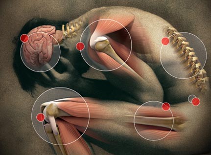
WELLNESS: Tendonitis: Its Causes, Symptoms and Treatments Tendons, which are flexible, tough and fibrous, connect your muscles to your bones. Muscles and tendons work together to exert a pulling force that helps you move properly. Tendonitis is an acute condition that occurs when injury to a tendon causes swelling and pain. It mainly affects your elbows, wrists, fingers, thighs, shoulders, knees and Achilles heels. There are two main tendonitis causes: sudden major injuries and repetitive movements over a time period while doing activities like: Gardening-Raking and shoveling-Carpentry-Cleaning or scrubbing-Painting-Throwing and pitching. Take note of these other factors that can increase a person's tendonitis risk: Age — Adults, especially those over 40 years old, are more injury-prone and have less-flexible and less-elastic tendons. Playing sports — Bowling, running, swimming, tennis, basketball, baseball or golf involve repetitive motions that can affect tendons. Health conditions — People with diabetes, rheumatoid arthritis, gout, psoriatic arthritis or thyroid disorders, those who experienced unusual reactions to medications, or people with hand or finger infections from cat or dog bites have a higher tendonitis risk. Body structure and posture — Tendonitis risk is higher with poor overall posture. Jobs or profession — Tendonitis is frequently seen in people whose jobs require them to stay in awkward positions, perform repeated actions, reach overhead, vibrate or forcefully exert their body. NOTE: Chiropractic helps.

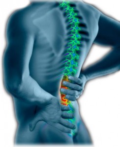
CHIROPRACTIC: Although doctors of chiropractic treat more than just back pain, many patients initially visit a chiropractor looking for relief from this pervasive condition. In fact, about 31 million Americans experience low back pain at any given time. Interesting Facts about Back Pain Worldwide, back pain is the single leading cause of disability, preventing many people from engaging in work as well as other everyday activities. Back pain is one of the most common reasons for missed work. One-half of all working Americans admit to having back pain symptoms each year.3Back pain accounts for more than 264 million lost work days in one year—that’s two work days for every full-time worker in the country. Experts estimate that up to 80% of the population will experience back pain at some time in their lives.5Back pain can affect people of all ages, from adolescents to the elderly. Back pain is the third most common reason for visits to the doctor’s office, behind skin disorders and osteoarthritis/joint disorders. Most cases of back pain are mechanical or non-organic—meaning they are not caused by serious conditions, such as inflammatory arthritis, infection, fracture or cancer. Most people with low back pain recover, however, reoccurrence is common and for a small percentage of people, the condition will become chronic and disabling. Worldwide, years lived with a disability caused by low back pain have increased by 54% between 1990 and 2015. Low-back pain costs Americans at least $50 billion in health care costs each year—add in lost wages and decreased productivity and that figure easily rises to more than $100 billion.

FUNNY BONE:. Q: What do you get when you cross a dinosaur with a pig? A: Jurrassic Pork.@@Q: What animal should you never play cards with? A: A cheetah!@@I went down the street to a 24-hour grocery store. When I got there, the guy was locking the front door. I said, "Hey! The sign says you're open 24 hours." He Said, "Yes, but not in a row!"@@Boy: The principal is so dumb! Girl: Do you know who I am? Boy: No... Girl: I am the principal's daughter! Boy: Do you know who I am? Girl: No... Boy: Good! *Walks away*@@Molecule 1: I just lost an electron. Molecule 2: Are you sure? Molecule 1: I’m positive.@@ When Magnesium and Oxygen started dating I was like, "O MG!"
Visit our web sites: keefeclinic.com & facebook.com/keefeclinic
Courtesy of:
John H. Keefe III, D.C.
(918) 663-1111

IN THE NEWS: Common Acid Reflux Medications Promote Chronic Liver Disease Approximately 10 percent of the general population take a proton pump inhibitor (PPI) drug to block stomach acid secretions and relieve symptoms of frequent heartburn, acid reflux and gastroesophageal reflux disease. That percentage can be as much as seven times higher for people with chronic liver disease. Researchers at University of California San Diego School of Medicine have discovered evidence in mice and humans that stomach (gastric) acid suppression alters specific gut bacteria in a way that promotes liver injury and progression of three types of chronic liver disease. The study is published October 10 in Nature Communications. "Our stomachs produce gastric acid to kill ingested microbes, and taking a medication to suppress gastric acid secretion can change the composition of the gut microbiome," said senior author Bernd Schnabl, MD, associate professor of gastroenterology at UC San Diego School of Medicine. "Since we found previously that the gut microbiome — the communities of bacteria and other microbes living there — can influence liver disease risk, we wondered what effect gastric acid suppression might have on the progression of chronic liver disease. We found that the absence of gastric acid promotes growth of Enterococcus bacteria in the intestines and translocation to the liver, where they exacerbate inflammation and worsen chronic liver disease." There are inexpensive and readily available alternatives to PPIs. However, even non-PPI-based antacids (e.g., Pepto-Bismol, Tums, or H2 blockers such as Tagamet and Zantac) still suppress gastric acid to a lesser degree. While these other types of antacids were not tested in this study, Schnabl said any medication that suppresses gastric acid effectively could cause changes in gut bacteria and thus potentially affect the progression of chronic liver disease. Alternatively, non-pharmacological methods for managing heartburn are an option for some patients, including losing weight and reducing intake of alcohol, caffeine, and fatty and spicy foods. An FDA report cautions against high doses or prolonged use of PPIs, because they’ve been shown to increase the risk of infection, bone fractures and dementia. But the danger doesn’t stop there. All acid stopping drugs (not just PPIs) inhibit nutrient absorption, promote bacterial overgrowth, reduce resistance to infection and increase the risk of cancer and other serious diseases.

CHIROPRACTIC: American Chiropractic Association (ACA) has announced a national health care observance October is National Chiropractic Month
DO YOU KNOW SOMEONE WITH A HEALTH PROBLEM OR IN PAIN?
INVITE YOUR FAMILY OR FRIENDS TO THE PARTY Who do you know who is hurting? Who do you know that you have been trying to get started at Keefe Clinic?
Now is the time to get your spouse or child started on the road to good health.
Complimentary DIAGNOSTIC WORK UP DURING OCTOBER
Complimentary initial exam and one X-ray. Tell someone today

WELLNESS: Best foods for good digestion Yogurt-has bacteria that is essentially good for your gut. You need to check their labels for "live and active culture" to reap the digestive benefits out of it. Banana-are very effective in treating gastric problems as they are helpful in restoring bowel function and can help treat diarrhea. They are rich in electrolytes and potassium which help in restoring good digestive health. Ginger-is a spice that has many benefits for digestive health. It can help cure motion sickness, nausea, vomiting, gas and loss of appetite. The ideal consumption would be 2 to 3 g every day. If you have more than that, it may cause heartburn. Beet root-are very good source of fiber, potassium and magnesium. They are an excellent cure for problems like constipation. Apple-are also rich in bacteria that are helpful in maintaining a good gut health. Avocado-are among the best sources of fiber and fruits. This helps in maintaining a mucosal lining in the gastrointestinal tract, which helps in the digestive process. Cod liver oil-source of the vitamin A and C, cod liver oil helps in maintaining a good digestive health. Blueberry-a very good source of fiber and vitamin C. They are loaded with cancer fighting properties. Kiwi-is loaded with minerals and nutrients which are very good for your gut health. It contains vitamin C and E, linolenic acid, magnesium, potassium, actinidin, fatty acids and pepsin which are good for your digestive health. Papaya-rich in papain which helps break down proteins in your stomach. Papaya also has anti-inflammatory properties which can help and soothing the stomach. It also helps relieve problems like food allergies and heartburn.
FUNNY BONE: TEACHER: Glenn, how do you spell crocodile? GLENN: K-R-O-K-O-D-I-a-L TEACHER: no, that’s wrong. GLENN: maybe it is wrong, but you asked me how I spell it.@@ I went to buy some camouflage trousers the other day but I couldn't find any.@@ If you jog in a jogging suit, lounge and a lounging pajamas, and smoke in a smoking jacket, why would anybody want to wear a windbreaker?@@ A police car pulled me over near the high school where I teach. As the officer asked me for my license and registration, my students began to drive past. Some honked their horns, others hooted, still, others stopped to admonish me for speeding. Finally, the officer asked me if I was a teacher at the school, and I told him I was. I think you’ve paid your debt to society, he said with a smile and left out given me a ticket.
Visit our web sites: keefeclinic.com & facebook.com/keefeclinic
Courtesy of:
John H. Keefe III, D.C.
(918) 663-1111

IN THE NEWS: Even More Evidence for the Link Between Alzheimer’s and Herpes In 1907, the German psychiatrist Alois Alzheimer published a description of a 50-year-old woman who suffered from memory problems, hallucinations, and delusions. In the woman’s brain, Alzheimer noticed unusual lumps, or “plaques,” which “were caused by the deposition of an unusual substance.” Eight decades later, the mystery substance was finally identified as a protein called amyloid beta. Though small, it can accumulate in large clusters that are somehow toxic to neurons. Those harmful plaques are one of the hallmarks of the disease that bears Alzheimer’s name. What amyloid beta normally does in the brain isn’t clear. Robert Moir, a neurologist at the MassGeneral Institute for Neurodegenerative Disease, says that many researchers have cast it as a villainous molecule with no beneficial function. “It’s just bad, bad, bad,” he says. “But it has become increasingly obvious that this isn’t true.” Moir thinks that amyloid beta has a more heroic role, as a foot soldier of our immune system. It protects neurons from infectious microbes—and from herpes viruses, in particular. William Eimer, a member of Moir’s team, demonstrated this protection by injecting the common herpes virus HSV–1 into the brains of two kinds of mice: normal rodents and ones that were genetically engineered to produce high levels of amyloid beta in their brains. The latter were better at resisting the viruses. Eimer then got similar results when he injected a different herpes virus, HHV–6, into human cells growing in a dish. Amyloid beta protects against these viruses by latching onto them in large numbers, imprisoning them in self-assembling cages. That’s typically a good thing, but Moir argues that if the process goes on for too long, it builds up to the problematic plaques of Alzheimer’s. According to him, amyloid beta is still at the heart of the Alzheimer’s story, but it isn’t the villain. “In our model, Alzheimer’s is caused by amyloid beta’s reaction to something else, and most likely some kind of infection” like herpes, he says. NOTE: Ask about nutrition for the immune system.
WELLNESS: Why Macadamias, Pecans and Walnuts Should Be on Your Shopping List Because macadamia nuts and pecans have high fat and low protein and carb levels, they are superior choices, particularly if you eat a ketogenic diet. Walnuts, despite having a slightly higher protein and carb content, are also an excellent choice mainly because new research suggests eating them may reduce your risk of Type 2 diabetes. You may not realize most nuts labeled as roasted have actually been fried in vegetable oil, often at high temperatures, putting you at risk of exposure to acrylamide, a possible carcinogen. When choosing nuts, look for high-quality nuts that are certified organic and presented in raw form. Avoid roasted or pasteurized nuts, as well as nuts coated in sugar or covered in milk chocolate. Contrary to what you may think, only dry-roasted nuts are truly roasted. Most nuts labeled and sold as roasted are actually fried in vegetable oil. You can tell this because the ingredient label will identify the type of oil used for frying. This practice is ill-advised for a few reasons:Most vegetable oils are unhealthy and contain an overabundance of omega-6 fats. In addition, some vegetable oils used to fry nuts, such as canola oil, are genetically engineered (GE). Roasting raises the potential for the formation of a possible carcinogen called acrylamide, which results from a chemical reaction between sugars in certain foods and an amino acid called asparagine. Acrylamide, which is best known as the “browning” on chips and french fries, has the potential to form on certain nuts when they are fried or roasted at temperatures above 250 degrees F (121 degrees C) for long cooking times. Vegetable oils heated to high temperatures can easily oxidize, promoting the formation of disease-causing free radicals. Nuts that oxidize can also become rancid and attract fungal mycotoxins. You can identify rancid nuts by a musty, stale or spoiled smell. Nuts roasted at high temperatures may contain lower amounts of antioxidants, vitamins and other nutrients.

CHIROPRACTIC: HELPING TMJ TMJ is the shortened name for temporomandibular joint disorders. It describes a group of disorders in which the connecting point between the jaw and the skull becomes painful and swollen. The cause of the disorder can vary widely, and in some instances, no known cause is ever discovered. What are the Symptoms? Symptoms of TMJ can vary widely from person to person, but some of the more typical problems are fairly common among most sufferers. These symptoms might include pain in the jaw, trouble chewing or talking, headaches, and neck pain. In some cases, the patient may also experience a bit of dizziness. Since other ailments can cause similar problems, it’s important to be seen by a professional. How Can a Chiropractor Help? Chiropractors may help with TMJ by alleviating tension and dysfunction in the spine. As chiropractors alleviate this dysfunction, it reduces the pressure on various nerves, which then alleviates pain associated with TMJ.
FUNNY BONE: Round like a shot... Going to bed the other night, I noticed people in my shed stealing things. I phoned the police but was told there was no one in the area to help. The Policeman said they would send someone over as soon as possible. I hung up. A minute later I rang again. 'Hello', I said, 'I called you a minute ago because there were people in my shed. You don't have to hurry now, because I've shot them.' Within five minutes there were half a dozen police cars in the area, plus helicopters and an armed response unit. They caught the burglars red-handed. One of the officers said: 'I thought you said you'd shot them.' To which I replied: 'I thought you said there was no one available.'@@ Why are iPhone chargers not called Apple Juice?!

It's really difficult for all of us to see when we are being programmed or manipulated through advertisement/propaganda. But pharmaceutical companies spend billions of dollars on conditioning the population to choose its products over other methods of treatment. And sometimes we’re in an automatic mode when we decide our choice in healthcare. This can be a problem for a number of reasons. The first thing to consider is if you are deciding to utilize a drug as your first option you might not understand the dangers of that approach.

Besides the over 2000 deaths per week due to the side effects of pharmaceutical drugs and the fact that medical care is a number one cause for death in the United States based on CDC statistics, there are other reasons to think twice about drugs being your first approach. Simply put drugs don't heal, drugs alter your perception of the condition. Let's take headaches, for example, most headaches are caused by misalignments in the upper neck (muscle tension) other causes could be toxicity problems, allergies, low blood sugar or hormonal problems. Which of these do you think an aspirin fixes? Did you know aspirin causes bleeding in the stomach, Excess Stomach Acid Secretion, Stomach Cramps, Blood coming from Anus, Decreased White Blood Cells? This is just a partial list of potential side effects. Again the aspirin doesn't treat any of the underlying causes for your condition.
Let's say your headaches were related to hormonal imbalances because of a developing ovarian cyst. You keep treating the headaches with an aspirin and your ovarian cyst keeps getting worse to the point that it might burst. Wouldn't it have been nice if the physician you went to wanted to understand the source of your headaches? But a common problem is if you're a physician and have powerful drugs that can mask symptoms then when a patient presents with certain symptoms you are just gonna write a prescription and consider it appropriate care.

As a chiropractic physician, we don't mask symptoms with drugs, we have to figure out why the symptom is there. Just to classify a headache is a tension headache or a migraine headache and then give the latest masking drug the pharmaceutical company has recommended, in my opinion, is not proper healthcare. Proper healthcare tries to determine the underlying cause for the condition and correct the cause. I'm not saying that can be done 100% of the time because science does not understand the body fully, I am saying that most the time it can be. The underlying cause of allergies is not an antihistamine deficiency it's an inappropriate functioning immune system. When you correct the immune system you correct the allergies. Acid reflux is not an antacid deficiency it is an imbalance somewhere within the digestive system. It needs to be found and corrected which might include changing your diet.
Back pain is not a muscle relaxer or anti-inflammatory deficiency disease. When you just treat the symptoms you allow the condition time to get worse. This is one of the reasons that chronic diseases are such a high prevalence in our society. When you chronically just treat symptoms over a lifetime you end up with a basket full of chronic conditions. And this is why many people live their last 10 or 15 years of life in a nursing home or a wheelchair suffering from multiple health problems. If you don't want to end up there you have to change the choices you make with your body. The fact is most drug therapy is inappropriate. Let me make this clear there is a time for drugs and there is a time for surgeries but they should be the last approach. Unfortunately, when you let certain conditions develop to the point of no return you eliminate a number of your options to get well.

Natural healthcare focuses on health, not disease. Disease care has led to horrible health statistics for countries that focus their attention there. When a type II diabetic is taking one, two or three different medications to try to control their blood sugar they are in fact shortening their lifespan and making chronic disease more likely. The sad thing is the vast majority of type II diabetics can be drug-free and healthy by some simple dietary and nutritional approaches. Lowering your blood sugar with drugs to force the sugar out of the body is not the same as correcting the blood sugar problem. Find the cause, correct the cause.

It is true that antibiotics can help clear acne in patients who suffer from it but it's also true that antibiotics will wreck havoc on your digestive tract and weaken your immune system. Is acne an antibiotic deficiency disease? The skin is just the expression of the health of the body. Acne has to do with body ecology, hormonal balance and sometimes food allergies. The chances are the use of antibiotics will make each of these issues worse. And has anybody heard of superbug infections? The overuse of antibiotics is the number one cause of death from superbug infections that have been growing every year for the past 25 years. Is there a place for antibiotics? There is but it's very rare. There are so many natural immune boosting techniques in natural healthcare that well over 90% of my patients never require any antibiotics in their life as long as they are following the approaches we recommend. Sure there are exceptions to every rule but in our society, the exception is the rule.

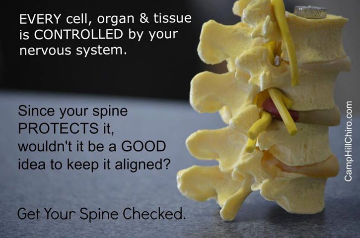
Do you want to be healthy? Drugs are not going to get you there. Drugs could save your life but the same drugs that saved your life could end your life if you keep taking them. Drugs are toxic, drugs are dangerous. If you want to be healthy then you have to practice a healthy lifestyle. Did you know your nervous system controlled and coordinated everything that happens in your body? That's why we focus on spinal health to maintain a proper nervous system function. That is one of the five laws of health that we encourage patients to follow. The second law would be the law of diet. Do you really believe you could eat Twinkies all day and stay healthy? Of course not. But just like the big Pharma, big food has programmed you into digging your own grave with your fork. You have to eat a diet that's consistent with your genetic makeup and actually has real food in it. Eating right is really not that hard with a little direction you can increase your energy levels, strengthen your cardiovascular systems and maintain proper blood sugar levels very easily. The third law is exercise. You don't have to run marathons, in fact, you can over-exercise to your detriment. Exercise is also related to your genetic makeup. Some people doing the wrong type of exercise can actually hurt themselves. People who have some type of moderate, regular exercise will be healthier than those who don't. Law four is a positive mental and spiritual attitude. Life can be hard you need to have coping skills in order to be successful. Spiritual disciplines have been shown by research to make us healthier and to allow us to live a longer life. In the fifth law is rest and relaxation.
None of these laws are really difficult to follow you just need to find yourself in a culture that encourages these. The pharmaceutical approach is not that culture. As nice as it is to have a fire department it's also a lot better if you never have to call them. And in most circumstances with a little prudence, those disasters can be avoided. One of the best ways to find yourself in the culture of better health is to choose natural healthcare. Making natural healthcare your first choice can prevent a number of disasters in your life and your families health. You still might be referred to a medical specialist from time to time but if your primary care is a physician in the natural healthcare field your outcomes would be greatly improved.

Are you locked in the matrix? Choose natural healthcare and choose a better life.
Mastic gum, also referred to as mastica,(click blue text to order) is the resin obtained from the Pistacia lentiscus tree, commonly sourced from the island of Chios in Greece. We also see this plant used historically throughout the Middle East and Northern Africa.
Mastic gum’s medicinal properties have been utilized for thousands of years for gastrointestinal ailments and related health concerns. These include the prevention of ulcers, ease of stomach discomfort, the killing off of bacteria, stubborn coughs, and teeth cleaning. Mastic gum is both antibacterial and antiviral. It has anti-inflammatory and anti-oxidant properties.
Mastic gum enjoyed a revival in the 1980s and 1990s when scientists discovered that it kills Helicobacter Pylori (H. Pylori). This infection affects the mouth, stomach, and intestines. Although this bacteria is present in billions of guts worldwide, when it becomes invasive it exacerbates or causes conditions like gastritis, peptic ulcers, stomach cancer, glaucoma, and Hashimoto’s.
Mastic gum has been used historically to treat cancers of breast, liver, spleen, and uterus. Modern science has shown the validity of its use in such cases, and added to this list its benefit to the colon and heart.
Mastic gum is also used to treat heartburn, gastroesophageal reflux disease (GERD), Crohn’s disease, chronic sore throat, herpes simplex, and to improve cholesterol levels.
Tangentially, mastic gum is used in traditional Greek, Turkish, and Arabic cooking.
How Mastica Works
Mastic is rich in terpenes. Terpenes are found in a variety of plants, giving them their unique scent and sometimes flavor. In this case, terpenes are the major organic compounds present in mastic gum’s resin that help to fight bacteria. The constituents are also believed to regulate and improve signaling between cells.
Many scientific papers state the effectiveness of mastic for various health conditions, yet state that the exact mechanism of action is still unknown.
When to Use Mastica
As aforementioned, the stomach’s pH, especially during digestion, is highly acidic. In a healthy stomach epithelial cells produce and secrete a thick layer of mucous to protect themselves from the acid and enzymes.
We require an acidic stomach not only to digest protein and other foods, but also to stimulate multiple digestive mechanisms, including the sphincter valve (lower esophageal sphincter [LES]) that connects the stomach to the esophagus (thus preventing heartburn and acid reflux), the liver that produces bile (which breaks down fat), the gall bladder that releases bile, and the ileocecal valve (between the large and small intestine) that when properly closed helps to prevent SIBO (small intestinal bacterial overgrowth). The proper stomach pH also helps to prevent the overgrowth of microorganisms and undigested food in the intestines (which leads to a systemic immune response, food sensitivities, inflammation, and autoimmune disease).
So while the stomach must be acidic during meals for digestion to work properly, we can work between meal times to rebuild the mucosal lining.
The burning feeling some patients feel in their stomachs during or after a meal, occurs when the acidic environment of the stomach comes into contact with damaged tissue. This burning feeling can become an ulcer, if not addressed.
Mastic gum can be taken in quantities between 1000 and 5000 mg. daily for many months, or longer, safely. In addition to killing harmful bacteria, it is speculated that it promotes new cell growth, thus rebuilding the epithelial cells that produce the mucosal lining.
One study recommends using mastica for many months to see a reduction in H. Pylori colonization levels. Due to emerging antibiotic resistance as well as the costliness of antibiotics, it’s considered an excellent alternative treatment. (I would add that antibiotics destroy the patient’s beneficial gut flora, largely foundational to good health.) Your doctor can determine if you have an overgrowth of H. Pylori through a blood test or with a urea breath test.
You can find mastic gum capsules here.(click blue text to order)
If it appeals to you to actually chew the resin’s “tears”, this is the gum (click blue text to order)that can be chewed! I believe there may be an additional benefit to choosing the gum option, as it kills bacteria in the mouth, stimulates the salivary glands (the first step in digestion), and acts much like oil pulling: our mouths help our bodies to detoxify! The gum is not sweet and has a mild pine flavor.
Ideally, patients can choose both options, as the capsules are easy to take between meals to help rebuild the mucosal lining and/or to kill bacterial overgrowth. And the gum, again between meals, is stimulating the entire alimentary canal and detoxifying the body.
A Healing Blend
If you’re someone who prefers fewer supplements but a multi-pronged approach, you might like this blend (click blue text to order)of mastic gum and deglycerized licorice, by Designs for Health. It also contains a few other excellent ingredients, necessary for gut healing, including vitamin C to help rebuild tissue, vitamin U (also known as the enzyme S methylmethionine, found in gentle, healing cabbage juice), and zinc for reducing inflammation.
Deglycyrrhized (DGL) Licorice
Many patients use deglycyrrhized licorice (click blue text to order)in conjunction with mastic gum. Licorice root has been used in Chinese medicine for centuries. Its many healing compounds have shown it to be anti-inflammatory, antibiotic, and rich in antioxidants.
When licorice root is sold as deglycyrrhized it’s been standardized to remove components that can cause side effects, making it safer universally. DGL should be used short term, or intermittently.
DGL can be used to help heal leaky gut. It soothes (increasing mucous production in the stomach), helps to prevent ulcers, works against nausea, stomach pain, and even helps to regulate cortisol levels, sometimes providing relief for adrenal fatigue. Licorice root is also anti-viral, great for the immune system.
This DGL (click blue text to order) comes in chewable form, first stimulating the salivary glands, and should be taken 20 minutes before meals.
Sources
https://www.ncbi.nlm.nih.gov/pubmed/19879118
https://www.ncbi.nlm.nih.gov/pmc/articles/PMC3127998/
Medicinal and Aromatic Plants of the Middle-East http://bit.ly/2qbkX0g
https://en.wikipedia.org/wiki/Terpene
https://www.ncbi.nlm.nih.gov/pubmed/16190616
https://bmcmedgenomics.biomedcentral.com/articles/10.1186/1755-8794-2-68
http://www.life-enhancement.com/magazine/article/770-mastic-is-more-than-an-antibacterial
http://www.life-enhancement.com/magazine/article/568-protect-your-stomach-from-deadly-bacteria
https://www.ncbi.nlm.nih.gov/pubmed/23162899
https://www.ncbi.nlm.nih.gov/pubmed/16884839
http://www.sciencedirect.com/science/article/pii/037851739400377H
https://www.ncbi.nlm.nih.gov/pubmed/21184804
Courtesy of:
John H. Keefe III, D.C.
(918) 663-1111

DIET: 43 Times More Vitamin C than an Orange, a Weapon Against the Flu Camu camu (Myrciaria dubia) is a small orange-red fruit, similar in size to that of a large cherry, with a tart flavor; the camu camu tree grows primarily in South America. Camu camu contains exceptionally high amounts of vitamin C — anywhere from 1,882 to 2,280 milligrams per 100 grams of fresh fruit. For comparison, acerola cherries typically contain around 1,678 mg of vitamin C per 100 grams. Vitamin C is an important immune booster and helps defend against viral attacks and inflammation. It’s also used by your body for wound healing, repairing and maintaining the health of your bones and teeth. Camu camu also contains a number of other valuable nutrients, including manganese, copper, carotenoids, flavonoids, essential amino acids (including valine, serine and leucine), gallic and ellagic acid, and fiber. Research suggests camu camu can help boost cognitive function and reduce your risk of dementia, facilitate muscle growth, aid digestion, lower your risk of obesity, Type 2 diabetes, cancer, macular degeneration and more.

IN THE NEWS: Untreatable 'super gonorrhea' case could be 'tip of the iceberg' The medical community is sounding the alarm after a man in the U.K. has become the first patient with a type of gonorrhea infection that is not responding to the antibiotics commonly used to cure it. Doctors are calling it the “worst-ever” case of the sexually transmitted disease, which hits approximately 78 million men and women every year. According to the BBC, the unidentified man had a regular partner but picked up the disease earlier this year after a sexual encounter with a woman in South East Asia. STDS HIT RECORD HIGH IN US, 2M CASES REPORTED IN 2016 "This is the first time a case has displayed such high-level resistance to both of these drugs and to most other commonly used antibiotics," said Dr. Gwenda Hughes, from Public Health England, to the BBC. If left untreated, gonorrhea can lead to pelvic inflammatory disease, ectopic pregnancy, stillbirths, severe eye infections in babies and infertility in both men and women. In the United States, there are approximately 300,000 reported cases of gonorrhea each year. But because infected people often have no symptoms, the actual number of cases is likely closer to 600,000, according to Reuters.

CHIROPRACTIC: Breaking the code of disease. One of the greatest rewards that a doctor of any type can have is helping someone recover their health. When we stopped taking care of ourselves like we should, it just a matter of time before we realize our mistake. Sometimes we don’t appreciate our health until we lose it. One of the tools that we have found very effective in “breaking the code of disease” is Vega testing. We have been able to help a multitude of patients recover their health after other methods have failed. One thing that’s important to realize is drugs primarily cover your symptoms. If you don’t fix the underlying cause and allow healing to take place, then you’re just giving disease time to develop into a more serious condition. I see that it has been some time since your last visit and I would like to encourage you to take a proactive stance concerning your health. Health is the best insurance you can have. Besides eating right for your body type, getting some type of regular movement (exercise), chiropractic adjustments and nutritional therapy are some of the most powerful tools in health recovery and maintenance. Are you noticing your performance and energy less than you would desire? Are you ignoring some reoccurring symptoms/pain or having issues with sleep or digestion? Are you having issues with brain fog or memory? Are you just not enjoying life like you use to? You need to call us today and let us unravel the issues affecting your well-being. Both you and your family would appreciate a healthier you. Call us today for an appointment, do something good for yourself.

FUNNY BONE: My dog used to chase people on a bike a lot. It got so bad, finally I had to take his bike away.@@ A woman walks into a library and asked if they had any books about paranoia. The librarian says "They're right behind you!"@@ Why did the old man fall in the well? Because he couldn't see that well.@@ I know a lot of jokes about unemployed people but none of them work.@@ My wife accused me of being immature. I told her to get out of my fort.@@ I just wrote a book on reverse psychology. Do *not* read it!@@ I used to think I was indecisive, but now I'm not too sure.
Visit our web sites: keefeclinic.com & facebook.com/keefeclinic
http://www.mdpi.com/2072-6643/10/5/604/htm
Open Access
Review
Inflammation, not Cholesterol, Is a Cause of Chronic Disease
Alexandros Tsoupras, Ronan Lordan and Ioannis Zabetakis *
Department of Biological Sciences, University of Limerick, V94 T9PX Limerick, Ireland
Received: 23 April 2018 / Accepted: 9 May 2018 / Published: 12 May 2018
Abstract
:
Since the Seven Countries Study, dietary cholesterol and the levels of serum cholesterol in relation to the development of chronic diseases have been somewhat demonised. However, the principles of the Mediterranean diet and relevant data linked to the examples of people living in the five blue zones demonstrate that the key to longevity and the prevention of chronic disease development is not the reduction of dietary or serum cholesterol but the control of systemic inflammation. In this review, we present all the relevant data that supports the view that it is inflammation induced by several factors, such as platelet-activating factor (PAF), that leads to the onset of cardiovascular diseases (CVD) rather than serum cholesterol. The key to reducing the incidence of CVD is to control the activities of PAF and other inflammatory mediators via diet, exercise, and healthy lifestyle choices. The relevant studies and data supporting these views are discussed in this review.
Keywords:
cardiovascular disease; atherosclerosis; inflammation; platelet-activating factor; oxidised lipoproteins; cholesterol; chronic diseases
1. Introduction
1.1. Biological Significance of Cholesterol—Circulating Blood Cholesterol
Cholesterol, an unsaturated alcohol of the steroid family, is essential for the normal function of all animal cells. It is also a fundamental element for the normal structural makeup and the fluidity of all cell membranes. Cholesterol interacts with phospholipid bilayers in the cell membrane and increases membrane packing. Cholesterol also takes part in signal transduction, intracellular transport, nerve conduction, and signalling pathways through lipid rafts and caveolae. Cholesterol has various other biological functions, i.e., it is a precursor molecule for several biochemical pathways such as the synthesis of vitamin D, steroid hormones (e.g., cortisol, aldosterone, and adrenal androgens), and sex hormones (e.g., testosterone, oestrogens, and progesterone). Cholesterol is also a constituent of bile salts, which are crucial constituents of digestion, as they facilitate the absorption of lipids, fats, and fat-soluble vitamins A, D, E, and K [
1].
Since cholesterol is mostly a lipophilic molecule, it does not dissolve well in blood. For this reason, it is packed into lipoproteins that are composed of a lipid core (which can contain cholesterol esters and triglycerides) and a hydrophilic outer membrane comprising phospholipids, apolipoprotein, and free cholesterol. This allows for the transport of the nonpolar lipid molecules such as cholesterol and triglycerides around the body through the blood to cells that require them. Plasma lipoproteins are separated into five major classes: chylomicrons, very-low-density lipoproteins (VLDL), intermediate-density lipoproteins (IDL), low-density lipoproteins (LDL), and high-density lipoproteins (HDL) [
1,
2].
Cholesterol can enter the blood through the digestion of dietary fat via chylomicrons. However, since cholesterol has an important role in cellular function, it can also be directly synthesised by each cell in the body. Notably, LDL particles are thought to act as a major transporter of cholesterol to the peripheral tissues, as at least two-thirds of circulating cholesterol resides in LDL. Conversely, HDL molecules are thought to do the opposite. They take excess cholesterol and return it to the liver for excretion [
1,
2].
Recent evidence suggests that dietary intake of cholesterol can influence plasma and serum levels, but not significantly. However, this is still subject to debate and further study [
3]. Plasma cholesterol levels along with the levels of LDL cholesterol, HDL cholesterol, and serum triglycerides are currently used as biomarkers of the so-called standard ‘lipid profile’ for each individual. The standard lipid profile has been widely used as a traditional biomarker, not only for cardiovascular health but also for other lipid-related abnormalities and disorders [
4].
1.2. Cholesterol Levels: Demonising a Risk Factor but Not the Causative Mechanisms of Chronic Diseases
Several modifiable and non-modifiable risk factors (genetic, environmental, nutrition, and lifestyle, etc.) are thought to influence the balance between health and disease by inducing mechanisms related to disease onset, development, and the manifestations of symptoms. The presence or coexistence of these risk factors seem to trigger underlying molecular and cellular mechanistic pathways that can lead to continuous chronic manifestations and the long-term loss of tissue homoeostasis and tissue dysfunction. These continuous chronic manifestations can develop over time before cellular disturbances manifest and cause tissue disorders, while, if not counterbalanced by our immune system and by specific preventive measures such as a healthy diet and lifestyle, the subsequent symptomatic disease finally appears, and medical treatment may be required to reduce the risk of mortality. Elucidating these molecular and cellular mechanistic pathways and acquiring the mechanistic evidence of the underlying multifactorial causes of a chronic disease can lead to suitable preventive targets against these diseases with fewer side effects, which is an ongoing difficult and demanding task. Such difficulties have misled the scientific and medical community to often and lightly extrapolate the easily acquired observed statistical and epidemiological correlations of traditional risk factors to several chronic diseases, towards matching these risk factors as the causative agents of these diseases.
According to the ‘cholesterol hypothesis’, high blood cholesterol is a major risk factor, while lowering cholesterol levels can reduce risk [
5]. Dyslipidaemias (i.e., hypercholesterolaemia or hyperlipidaemia) are abnormalities of lipid metabolism characterised by increased circulating levels of serum total cholesterol, LDL cholesterol, triglycerides, and decreased levels of serum HDL cholesterol. High levels of LDL cholesterol and non-HDL cholesterol have been associated with cardiovascular risk, while other cholesterol-related serum markers, such as the small dense LDL cholesterol, lipoprotein(a), and HDL particle measurements, have been proposed as additional significant biomarkers for CVD risk factors to add to the standard lipid profile [
6]. HDL cholesterol has been considered as the atheroprotective ‘good’ cholesterol because of its strong inverse correlation with the progression of CVD [
7]; however, it is the functionality of HDL cholesterol, rather than its concentration that is more important for the preventative qualities of HDL cholesterol in CVD. In general, dyslipidaemias have been ranked as significant modifiable risk factors contributing to prevalence and severity of several chronic diseases including aging, hypertension, diabetes, and CVD. High serum levels of these lipids have been associated with an increased risk of developing atherosclerosis [
8].
Furthermore, dyslipidaemias have been characterised by several studies not only as a risk factor but as a “well-established and prominent cause” of cardiovascular morbidity and mortality worldwide [
9]. Even though such an extrapolation is not adequate, it was, however, not surprising that this was made, because since the term arteriosclerosis was first introduced by pioneering pathologists of the 19th century, it has long been believed that atherosclerosis merely involved the passive accumulation of cholesterol into the arterial walls for the formation of foam cells. This process was considered the hallmark of atherosclerotic lesions and subsequent CVD. Moreover, one-sided interpretations of several epidemiological studies, such as the Seven Countries Study (SCS), have highlighted outcomes that mostly concerned correlations between saturated fat intake, fasting blood cholesterol concentrations, and coronary heart disease mortality [
10,
11,
12,
13]. Such epidemiological correlations between dyslipidaemias and atherosclerosis led to the characterisation of atherosclerosis as primarily a lipid disorder, and the “lipid hypothesis” was formed, which would dominate thinking for much of the 20th century.
In the clinical setting, in order to address the lipid hypothesis, the levels of cholesterol related plasma lipoproteins and triglycerides (lipid profile) have been used as traditional biomarkers for cardiovascular risk, but also for dietary and treatment guideline designs [
5]. Dietary and medical guidelines have focused on the reduction of cholesterol and lipid levels as the best way to prevent chronic diseases such as CVD [
5,
9]. Such guidelines suggest the application of statin therapies in order to reduce the levels of cholesterol (through inhibition of cholesterol synthesis by HMG-CoA reductase inhibitors); however, numerous side effects have been reported, including the development of other chronic diseases such as diabetes mellitus [
14]. Moreover, specific dietary strategies for reducing cholesterol intake are the mainstay of management in most cases of dyslipidaemia, prior to, or simultaneously with, the initiation of a lipid lowering agent [
9]. Dietary fats, cholesterol, and the levels of serum cholesterol in relation to the development of CVD have been somewhat demonised.
On the other hand, since cholesterol is an essential biomolecule for the normal function of all our cells, an emerging question has recently surfaced: “how much do we need to lower the levels of cholesterol”? Furthermore, given the fact that cholesterol plays a crucial role in several of our cellular and tissue mechanisms, it is not surprising that there are several consequences due to the aggressive reduction of cholesterol levels in the body, which has been common practice over the last few decades. In addition, targeting cholesterol and fat intake by introducing diets with low-fat products and by reducing the intake of high-fat foods can lead to less absorption and lower bioavailability of other lipids containing high value nutrients, such as several lipid soluble vitamins (especially vitamin D) and other lipid molecules. Such lipids have exhibited a plethora of beneficial bioactivities, not only related to reducing the risk of chronic diseases but also through a wide range of important bio-functionalities and anti-inflammatory properties [
3]. Therefore, lower cholesterol levels do not equate to better health, or to lower risk of chronic diseases such as CVD. Homeostasis must be maintained, even with regard to cholesterol, both HDL and LDL [
15].
Moreover, recent systematic reviews and meta-analyses have started to question the validity of the lipid hypothesis, as there is lack of an association or an inverse association between LDL cholesterol and both all-cause and CVD mortality in the elderly [
15] and several cancers such as lung, prostate, and breast cancer [
16,
17,
18]. Such studies provide the rationale for more research about the causes (and not only the risk factors) of chronic diseases such as atherosclerosis, CVD, and cancer, but also for a re-evaluation of the guidelines for cardiovascular prevention, in particular because the benefits of statin treatments have been exaggerated [
15].
Statistical and epidemiological extrapolations often lack fully clarified biochemical mechanistic evidence, while associations and correlations do not necessarily mean causation. In addition, a follow-up by systematic reviews and meta-analyses often present contradictory outcomes against the initial results that were introduced by early stage epidemiological studies lacking consistency, biological gradient, and coherence. Thus, such extrapolations can lead to one-sided, premature targeting of risk factors accompanied with consequences, often without the desirable outcomes. Targeting a risk factor such as high serum cholesterol may decrease the probabilities for a disease, but usually cannot prevent the causation of chronic diseases.
1.3. Revisiting the Lipid Hypothesis: Outcomes of the Mediterranean Diet against Inflammation
Previous epidemiological and observational studies, such as the SCS in which the lipid hypothesis was mostly based, have been re-evaluated. For example, even though within the SCS the strength of the association between serum cholesterol and cardiovascular mortality were similar in different cultures, the absolute risks differed substantially. Kromhout reported that at a serum cholesterol level of 200 mg/dL, the 25-year cardiovascular mortality rate was five times higher in the Northern European populations of the SCS compared to the Southern Mediterranean populations [
19], and thus the relations between diet, serum cholesterol, and cardiovascular mortality are more complex than originally thought. This is because it is not only dietary cholesterol involved, but other lipids and antioxidants may play a role in the onset and prevention of atherosclerosis [
19]. Such a low prevalence of cardiovascular mortality in the Mediterranean cohorts of SCS is now attributed to their lifestyle and especially to their dietary habits, namely the traditional Mediterranean diet (Med-diet) [
10,
20]. A common feature of the diet amongst populations in the Mediterranean is a relatively high dietary intake of vegetables, fruits, legumes, whole grains, monounsaturated fats, and nuts, followed by moderate consumption of fish, dairy products (mainly cheese and yogurt), alcohol, and low consumption of red and processed meats [
21].
The major outcomes of the SCS and other similar epidemiological studies (i.e., studies trying to decipher the ‘French Paradox’ [
22]) concerning the protective effects of dietary patterns, such as the Med-diet against chronic diseases, were initially either neglected or misinterpreted. CVD and cardiovascular mortality occurred in much relatively lower rates in the Southern European populations (i.e., Italy and Greece) despite a rather high dietary intake of saturated fats and cholesterol [
10,
20,
23]. A recent systematic review and meta-analysis revealed that Med-diet can actually reduce the incidence of cardiovascular events, breast cancer, and type II diabetes mellitus, without any restriction on fat intake [
24].
Over the last 2 years there has been a significant number of studies referring to adoption of the Med-diet pattern and its associated beneficial outcomes in a plethora of several chronic diseases that are either directly or indirectly related to inflammation. These studies refer to heart failure, CVD [
25], cancer [
26,
27], obesity [
28], metabolic syndrome [
29,
30,
31], diabetes [
31,
32,
33,
34], and other subsequent manifestations such as diabetic retinopathy [
35], asthma [
36], autoimmune diseases such as rheumatoid arthritis [
37], incident frailty risk [
38], non-alcoholic fatty liver disease [
39,
40], inflammatory bowel disease [
41], cognitive health, the risk of Alzheimer’s disease and dementia [
42,
43,
44], and age-related macular degeneration [
45].
In addition, the Med-diet has also been associated with beneficial outcomes, even in secondary CVD prevention [
46]. When patients suffering from CVD or diabetes follow the Mediterranean dietary pattern, the incidence of recurrent myocardial infarction and cerebrovascular events is reduced. The protective effect of this dietary pattern can be maintained for up to four years after the first infarction (Lyon Diet Heart Study) [
47]. Moreover, in contrast to the contradictions of lipid hypothesis and mortality in elderly people [
15], the HALE project has also shown that individuals aged 70 to 90 years following a Med-diet and healthy lifestyle have a 50% lower rate of all-cause and cause-specific mortality [
48]. Followers of the Med-diet are also less likely to suffer sudden cardiac death and age-related cognitive decline [
49].
The inverse association between Med-diet and all causes of diseases and cardiovascular-mortality has been attributed to several of its pleiotropic protective effects. For instance, the Med-diet can beneficially influence several risk factors such as lowering BMI, blood pressure, reducing insulin resistance, reducing lipid levels (i.e., the ratio of cholesterol/HDL cholesterol), and improving HDL-cholesterol functionality [
50,
51,
52,
53,
54]. However, the main beneficial impact of Med-diet is on the improvement of endothelial function and the decrease of the inflammatory milieu, inflammation-related mediators, biomarkers such as platelet-activating factor (PAF), and several cytokines. It is also suggested that there is an improvement of oxidative stress, with lower concentrations of oxidised LDL and improved apolipoprotein profiles, and, finally, there is evidence of beneficial effects against platelet aggregation and blood coagulation [
3,
55,
56,
57,
58].
The overall outcomes and beneficial effects of Med-diet have radically shifted the attention from the lipid-centric model that is characterised by the desired reduction of cholesterol levels to more effective targeting against the factual causative factors of chronic diseases, which are inflammation and its related manifestations. Prevention is key to reducing global mortality due to chronic diseases such as CVD; therefore, it is imperative to separate the underlying causes and processes of the disease from the risk factors and symptoms of disease. The clarification of the key roles and interplay of various cells, inflammatory mediators, and pathways during chronic inflammatory manifestations related to the onset of several chronic diseases is of great importance and may lead to a plethora of novel potential targets for fine-tuning of the inflammatory response during the chronic smouldering of inflammation that characterises these disorders.
2. Re-Discovering Chronic Inflammation as the Cause for Chronic Diseases
Inflammation is a physiological reaction of the innate immune system that maintains a constant internal milieu while being exposed to continuously changing environmental pressures, irrespective of whether the initial causes originate from mechanical, physical, chemical, infectious, immunological, or reactive natural traumatic injury or metabolic dysfunction. The inflammatory response aims to reduce the agent that causes tissue injury and/or minimise these effects, to induce appropriate wound healing and to restore tissue homeostasis. Inflammatory responses are initiated by innate sensing mechanisms that detect the presence of microbial infection, stressed or dying cells, loss of cellular integrity, barrier breach, etc. A cascade of inflammatory pathways and mechanistic effects is supposedly well-orchestrated by the immune system in order to eradicate the causative agent.
Several immune cells can change their number, morphology, and nature depending on the stage and type of inflammation. Biochemically, inflammation is denoted by a local increase of numerous tissue hormones, transmitters, complement components, cytokines, and lipid mediators such as PAF and eicosanoids. Most of these products are autacoids that are synthesised at the site of inflammation in order to resolve the inflammatory process by removing or inhibiting the actions of the triggering agent [
8]. Provided that the immune response succeeds in eliminating the infectious agent or to repair the initial tissue injury, the inflammatory process will be terminated in a timely fashion and thus only affects tissue function transiently.
However, in cases where the inflammation fails to resolve due to the persistence of the triggering agent or due to unsuccessful repair of the initial tissue injury or dysfunction, a sustained underlying inflammatory process develops, leading to further tissue dysfunction and detrimental consequences. Several traditional and emerging risk factors are thought to influence our health and, especially, inflammation-related chronic diseases, by their interrelation with underlying molecular and cellular manifestations that result in chronic inflammatory responses leading to the loss of tissue homoeostasis and dysfunction. Apart from dyslipidaemias, other well-established risk factors include hypertension, diabetes, smoking, excessive food intake, previous infections (influenza, oral pathogens) or underlying autoimmune diseases such as lupus or rheumatoid arthritis, pollution, and genetic abnormalities [
59]. It is now well established that a common junction of such risk factors is chronic and unresolved inflammatory manifestations. Inflammation that causes endothelial dysfunction seems to be the key causative underlying mechanistic player, at the molecular and cellular level, for the onset and development of subsequent inflammation-related chronic disorders such as atherosclerosis and subsequent CVD, ischemic and renal disorders, cancer metastasis, diabetes, infections, and comorbidities [
8,
57,
58,
59,
60,
61,
62].
For example, in cases of dyslipidaemia, increased cholesterol levels are not the causative agent or the underlying biochemical mechanism responsible for endothelial dysfunction and atherosclerosis development. The accumulation of excess plasma LDL cholesterol is addressed by the innate immune system as an undesired event. Therefore, an inflammatory response at the endothelial wall is promoted to reduce the threat by the removal of excess LDL and oxidised-LDL (Ox-LDL) cholesterol from the blood stream to the subendothelium, where they are engulfed by comigrated monocytes for final removal [
63,
64]. During chronic inflammatory diseases, inflammation and infections can also induce a variety of alterations in lipid metabolism, including decreases in serum HDL cholesterol, increases in triglycerides, lipoprotein(a), and LDL levels. These changes of the lipid levels may initially dampen inflammation or fight infection; however, the sustained inflammation can contribute to the increased risk of atherosclerosis [
65]. In addition to affecting serum lipid levels, inflammation also adversely effects lipoprotein function; LDL is more easily oxidised, as the ability of HDL to prevent the oxidation of LDL is diminished, while several steps in the reverse cholesterol transport pathway are also adversely affected during inflammation. The greater the severity of the underlying inflammatory disease, the more consistently these abnormalities in lipids and lipoproteins are observed [
65]. Thus, it is not serum cholesterol and lipoproteins that influence the endothelium but the inflammatory response that affects the well integrity and functionality of the endothelium.
Apart from the effects of inflammation on plasma lipids, it is now well established that more important soluble and cellular immune factors associated with chronic inflammation can promote inflammation-related endothelial dysfunction and atherogenesis, either during dyslipidaemia or independently of dyslipidaemia [
66]. Even though atherosclerosis and CVD were previously viewed as lipid storage disorders, we now recognise that inflammation drives much of endothelial dysfunction and mechanisms of clinical complications with these diseases and related comorbidities, such as sepsis [
67,
68], human immunodeficiency virus (HIV) infection [
69,
70,
71,
72,
73,
74], periodontal diseases [
75,
76,
77], kidney disorders [
78,
79,
80,
81], healthy ageing, and inflammatory autoimmune diseases such as systemic lupus erythematosus and rheumatoid arthritis, independently of traditional cardiovascular risk factors such as serum lipid levels [
66,
82,
83].
Inflammation plays a key role in all stages of the formation of vascular lesions maintained and exacerbated by several risk factors such as unhealthy diet and lifestyle, smoking, hyperlipidaemia/hypercholesterolaemia, hypertension, autoimmune diseases, etc. The consequence of chronic inflammation is endothelial dysfunction that sets in, and we can define it as an integrated marker of the damage to arterial walls by classic risk factors. Endothelial dysfunction is usually characterised by an inflammation-related milieu acting on leukocytes and endothelial cells, through an interplay with other immune cells such as T lymphocytes, mast cells, dendritic cells (DC), and platelets [
57,
58,
66,
84,
85]. The orchestrated overexpression and increased production of pro-inflammatory cytokines occurs, including interleukin-6 (IL-6), tumour necrosis factor (TNF) and its receptor, high-sensitivity C-reactive protein (hsCRP), type I interferons (IFN-α, IFN-β), adhesion molecules, chemokines, and lipid inflammatory mediators such as PAF and eicosanoids. Other linked events include the increased generation of reactive oxygen species (ROS), the increased oxidation of LDL cholesterol, and the reduction of protective nitric oxide levels.
Therefore, the mechanistic pathways and key players implicated in the inflammatory crosstalk taking place throughout the onset, development, and progression of chronic diseases is of great importance, in order to unravel putative preventive and therapeutic targets with less side effects. The inverse effects of the Med-diet with chronic diseases is mostly related to the pleiotropic effects and interplay of its food constituents on all these inflammation-related pathways; following a Mediterranean dietary pattern leads to the reduction of several inflammatory mediators and biomarkers related to the endothelial functionality, such as decreases in hsCRP, IL-6, and intracellular adhesion molecule-1 (ICAM-1) [
27].
3. The Role of PAF in Chronic Diseases and the Beneficial Effects of the Mediterranean Diet
3.1. PAF Structure, Activities, and Metabolism: The Role of PAF
3.1.1. PAF Structure and Physiological Roles
PAF is a potent lipid inflammatory mediator with pleiotropic effects that are implicated in several chronic diseases [
57]. The classic PAF molecule is characterised by an alkyl ether linkage at the
sn-1 position, an acetyl group at the
sn-2 position, and a phosphocholine group at the
sn-3 position of glycerol backbone (1-O-alkyl-2-
sn-acetyl-glycero-3-phosphocholine, [
86]) (A). These three structural features are all equally important requisites for the optimal biological activity of PAF, mediated by its stereospecific binding to its specific receptor [
87,
88]. Because of the ether linkage at the
sn-1 position, the classic molecule of PAF is an unusual lipid, as such moieties are not common in animals, nor is it common to find the acetic acid esterified directly to glycerol at the
sn-2 position. Thus, it seems that PAF was chosen by evolution to participate in specific functions in several of our cells, tissues, organs, and throughout the body. PAF was the first intact phospholipid known to have messenger functions by binding to a specific receptor on the cell membrane, and not simply via physicochemical effects on the plasma membrane of the target cell [
79].
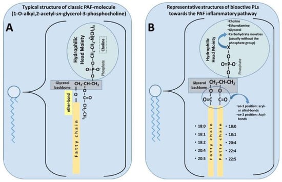
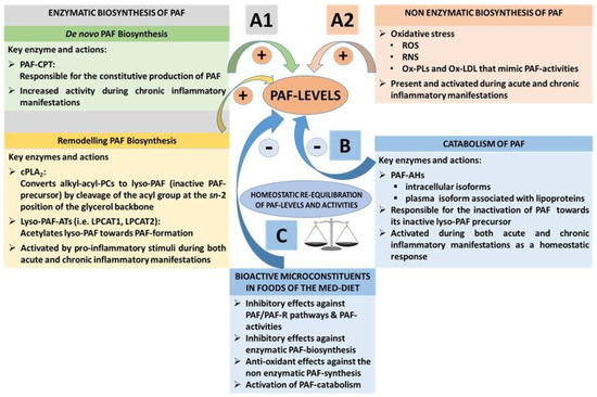
Figure 1. (
A) Typical structure of classic platelet-activating factor (PAF) molecule [
86]. (
B) Representative structures of bioactive polar lipids (PL) towards the PAF inflammatory pathways (
B), which have been identified in several foods of the Mediterranean diet [
56].
Lately, the term ‘PAF family’ has been proposed to include every other phospholipid molecule called PAF-like molecules, which have similar structures to those of the classic PAF molecule, and they exhibit similar bioactivities [
89]. However, such PAF-like moieties are usually less potent than PAF by several orders of magnitude, i.e., increasing the chain length beyond 3 carbons at the
sn-2 position decreases its biological potency; likewise, altering the polar group at
sn-3 position decreases the potency of the molecule. The molecular composition of PAF varies depending on different species and cell types. Related PAF-like lipids include, for example, the acyl-phosphatidylcholine-PAFs (with a short chain acyl group at the
sn-2 position), ethanolamine-PAFs, inositol-PAFs, oxidised alkyl-acyl phosphatidyl glycerophosphocholines [
90,
91], and hydroxyl-alkyl acyl phospholipids [
76,
77].
PAF, in general, play a vital role in various physiological processes such as mediation of normal inflammatory responses, regulation of blood circulation and pressure, regulation of coagulation responses, glycogen degradation, brain function, reproduction, foetal implantation, lung maturation, initiation of parturition, and exocrine gland functions [
92]. However, PAF can be regarded as both a friend, since it is presumed to have evolved as part of a protective mechanism in the innate host defence system, but also as a foe, because of its involvement in uncontrolled inflammation-related pathological conditions [
93]. When present in excess, PAF has been implicated in the pathogenesis of several inflammation-related chronic disorders [
57]; thus, its synthesis, distribution, and degradation are all strictly controlled, as would be predictable for such a potent molecule with a wide range of diverse actions.
3.1.2. The PAF/PAF-Receptor Signalling Pathways
PAF and PAF-like molecules act through their binding to a unique G-protein coupled seven transmembrane receptors, called the PAF-receptor (PAF-R) [
87,
88]. Species identity, differentiated by heterogeneity in linkage, degree of unsaturation, and carbon chain length of the alkyl or acyl chains at the
sn-1 and
sn-2 position, partially dictates signalling specificity by eliciting various signal transduction pathways following PAF-R activation [
94,
95]. The PAF-R is constitutively present on platelets, leukocytes, and endothelial cells, and further expression may be induced by appropriate stimuli. PAF-R is highly expressed by cells within the innate immune and cardiovascular systems [
96], pointing to a role for PAF and PAF-like molecules as pleiotropic communicators in plasma [
97].
Ligand binding (PAF and/or PAF-like molecules) to the PAF-R subsequently triggers multiple intracellular signalling pathways and gene-expressions, depending on the target cell and PAF levels (concentration) in blood or tissue [
87,
88,
89,
98] ( (A1–A3)). For example, activation of the PAF-R signalling initiates (through a Gq-linked mechanism) PLCβ-mediated hydrolysis of PIP2 to produce IP3 and DAG, leading to transient elevation of cytosolic Ca
2+ released from intracellular stores and activation of PKC. The rise in Ca
2+ also activates cPLA2α, leading to the release of arachidonic acid (AA) and lysophosphatides, which can serve as substrates for further synthesis of eicosanoids and PAF, respectively. In addition, signalling through Gi-linked PAF-R inhibits the conversion of ATP to cAMP by adenylate cyclase, thus preventing the activation of PKA and related signalling events.
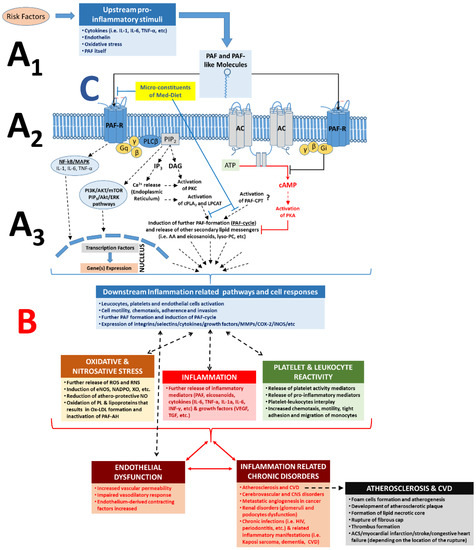
Figure 2. Role of PAF, PAF-R, and its related pathways in the inflammatory cascades and in the pathogenesis of inflammation-related chronic disorders; increased PAF levels by pro-inflammatory stimuli and binding of PAF on its receptor, PAF-R, on the membranes of several cell types can lead to intracellular cascades and a PAF cycle-related amplification of the initial stimuli (
A) and in numerous cell responses according to each cell type (
B), which can lead to endothelial dysfunction and the onset and progression of inflammation-related chronic diseases. A1. Several risk factors and related upstream pro-inflammatory stimuli trigger formation of PAF and PAF-like molecules (i.e., oxidised phospholipids) and expression of PAF-R. A2. Binding of PAF/PAF-like molecules on PAF-R promote several inflammation-related intracellular pathways; activation of the PAF-R signalling initiates (through a Gq-linked mechanism) PLCβ-mediated hydrolysis of PIP
2 to produce IP
3 and DAG, leading to transient elevation of cytosolic Ca
2+ released from intracellular stores and activation of PKC. The rise in Ca
2+ also activates cPLA
2α, leading to the release of AA and lysophosphatides, which can serve as substrates for further synthesis of eicosanoids and PAF, respectively. Signalling through Gi-linked PAF-R inhibits the conversion of ATP to cAMP by adenylate cyclase, in this way preventing the activation of PKA and related anti-inflammatory signalling events. A3. Activation of the PAF/PAF-R intracellular pathways leads to the activation of cPLA
2 and PAF biosynthetic enzymes (LPCAT) for further formation of PAF and other lipid second messengers, thus creating a PAF cycle and further amplification of the initial inflammatory stimuli, while expression of genes involved in inflammatory manifestations (such as genes of several cytokines, integrins, selectins, metalloproteinase, several enzymes for eicosanoids, and ROS, etc.) is also induced. The pathways inducing the PAF-CPT-related synthesis of PAF are not fully elucidated. B. Increased PAF levels at the site of inflammation and ligand binding (PAF and/or oxidised phospholipids binding) on PAF-R can promote a broad spectrum of PAF effects depending on the cell type and tissue, which is achieved through the production and release of various downstream mediators, such as PAF itself and several other mediators of inflammation such as eicosanoids, cytokines (i.e., TNF-α, IL-1α, IL-6, IL-8, INF-γ, etc.), growth factors (i.e., VEGF, IGF, TGF), ROS, and RNS, but also through the expression of selectins and integrins (i.e., ICAM, VCAM, P-Selectin, E-Selectin) in the membranes of activated cells. Thus, increased downstream mediators, PAF levels, and the subsequent further activation of the PAF/PAF-R pathways promotes the activation and aggregation of platelets and leukocytes, activation of endothelial cells, leukocyte adherence, motility, chemotaxis, invasion, migration, and subsequent endothelial dysfunction, thus stimulating the onset and development of inflammation-related chronic diseases and disorders. C. Microconstituents of several foods of the Mediterranean diet have been found to beneficially inhibit the PAF/PAF-R pathways and PAF synthesis towards homeostatic re-equilibration of PAF levels and activities [
57]. PAF: platelet-activating factor; PAF-R: G-protein-coupled PAF-receptor; AC: adenylate cyclase; NF-kB: nuclear factor-kappa light-chain-enhancer of activated B cells; MAPK: mitogen activated protein kinase; ERK: extracellular signal-regulated kinases; Akt: protein kinase B; PI3K: phosphatidylinositol 3-kinase; mTOR: mechanistic target of rapamycin; DAG: diacylglycerol; AA: arachidonic acid; cPLA
2: cytosolic phospholipase A
2; PKC: protein kinase C; PKA: protein kinase A; LPCAT: acetyl-CoA: lyso-PAF acetyltransferases; PAF-CPT: dithiothreitol l-insensitive CDP-choline: 1-alkyl-2-acetyl-
sn-glycerol cholinephosphotransferase; ATP: adenosine triphosphoric acid; cAMP: cyclic adenosine monophosphate; PLC: phospholipase C; MMP: metalloproteinase; COX: cyclooxygenase; iNOS: nitric oxide synthase; eNOS: endothelial nitric oxide synthase; ROS: reactive oxygen species; RNS: reactive nitrogen species; NADPO: nicotinamide-adenine dinucleotide phosphate oxidase; XO: xanthine oxidase; IL-6: interleukin-6; IL-1: interleukin-1; TNFα: tumour necrosis factor-α; ACS: acute coronary syndrome; VEGF: vascular endothelial growth factor; PL: phospholipids; CVD: cardiovascular diseases; CNS: central nervous system.
Signalling through other pathways is also amplified by the PAF/PAF-R pathway activation, since inhibition of PAF synthesis or PAF-R blockade significantly attenuates signalling through apparently unrelated pathways, suggesting a critical role for PAF/PAF-R action as a co-stimulatory signal. For example, many VEGF-directed effects on vascular endothelium require PAF synthesis [
57]. Nevertheless, the activation of the PAF/PAF-R pathway further triggers the activation and aggregation of platelets and leukocytes and promotes leukocyte and platelet adherence, motility, chemotaxis, invasion, migration, ROS generation, and further PAF formation () [
89,
98].
3.1.3. PAF Levels Result from Enzymatic Biosynthesis, Non-Enzymatic Oxidative Synthesis, and Enzymatic Catabolism
Under normal circumstances, homeostatic levels of PAF present in plasma and biological tissue seem to be regulated by a balance of its biosynthetic and catabolic enzymatic pathways [
57]. PAF is synthesised throughout the body by the specific stimulation of various cell types such as platelets, macrophages, monocytes, eosinophils, basophils, and endothelial cells. PAF is mostly produced in the blood, lungs, kidney, myocardium, brain, liver, skin, saliva, retina, uterus, and embryo [
56,
99,
100]. Two enzymatic pathways by which PAF is biosynthesised in the body are the ‘remodelling’ and the ‘
de novo’ pathways ((A1)).
Figure 3. PAF levels result from enzymatic biosynthesis, non-enzymatic oxidative synthesis, and enzymatic catabolism, while bioactive microconstituents of the Med-diet beneficially affect these pathways. (A1) The enzymatic biosynthesis of PAF contributes to basal PAF levels or a periodic increase of PAF levels during normal inflammatory responses, while during unresolved and chronic inflammatory manifestations, the enzymatic biosynthesis of PAF is responsible for pathologically increased PAF levels through a continuous induction of the PAF cycle; (A2) Non-enzymatic synthesis of PAF occurs during oxidative stress, increasing ROS and RNS and inducing the synthesis of PAF and PAF-like molecules. When Ox-LDL is produced, PAF-like molecules mimic the activities of PAF. These pathways are not regulated enzymatically; (B) Catabolism of PAF is enzymatically regulated by PAF-AH. PAF catabolism is activated during both acute and chronic inflammatory manifestations and inactivates both PAF and PAF-like molecules; (C) Bioactive microconstituents present in foods of the Med-diet (i.e., polar lipids) have demonstrated beneficial outcomes by inducing homeostatic equilibration of PAF levels and activities through the Inhibition of the PAF/PAF-R pathways and modulation of the PAF anabolic and catabolic enzymes. PAF: platelet-activating factor; PAF-R: G-protein coupled PAF-receptor; PAF-CPT: dithiothreitol l-insensitive CDP-choline: 1-alkyl-2-acetyl-sn-glycerol cholinephosphotransferase; Lyso-PAF-ATs (LPCAT1, LPCAT2): acetyl-CoA: lyso-PAF acetyltransferases; cPLA2: cytoplasmic phospholipase A2; PAF-AH: PAF-acetylhydrolase; PC: Phosphatidylcholine; ROS: reactive oxygen species; RNS: reactive nitrogen species; LDL: low-density lipoprotein; Ox-LDL: oxidised-LDL; Med-diet: Mediterranean diet.
The remodelling enzymatic pathway of PAF biosynthesis involves remodelling of a membrane lipid constituent (a long-chain fatty acyl residue in
sn-2 is replaced with an acetyl residue), and it has been proposed that this pathway is periodically involved in the acute pro-inflammatory production of PAF under activation of several cells during inflammation [
101]. More specifically, the action of cytoplasmic phospholipase A
2(PLA
2) yields a precursor of PAF called lyso-PAF (1-O-alkyl-
sn-glyceryl-3-phosphorylcholine), which is then acetylated by at least two isoforms of acetyl-CoA: lyso-PAF acetyltransferases, namely, LPCAT1 and LPCAT2 (lyso-PAF AT), leading to the formation of PAF [
102]. LPCAT2 is highly expressed in inflammatory cells, and, depending upon the inflammatory stimulus used to activate the cells, PAF is produced within seconds, minutes, or hours following stimulation. In addition, PAF itself can act as an inflammatory signal, and the binding of PAF to its receptor on inflammatory cells can promote the very rapid (within 30 s) production of PAF; PAF-induced, protein kinase, Cα-mediated phosphorylation of LPCAT2 enhances enzymatic activity, leading to the vary rapid production of PAF. Thus, a PAF cycle can consistently induce increased PAF levels and subsequent inflammatory cascades ( and )
The
de novo enzymatic pathway of PAF biosynthesis is similar but distinct to the biosynthesis of phosphatidylcholine, since a phosphocholine function is transferred to alkyl acetyl glycerol. This pathway has been initially reported as the pathway responsible for the constitutive production of PAF basal levels. A key step in this route is the conversion of 1-O-alkyl-2-
sn-acetyl-glycerol to PAF by a specific dithiothreitol l-insensitive CDP-choline: 1-alkyl-2-acetyl-
sn-glycerol cholinephosphotransferase (PAF-CPT) [
57,
81]. Interestingly, apart from the remodelling pathway, which is always activated in both acute and chronic inflammation, the key enzyme of the ‘
de novo pathway, PAF-CPT, seems to be more active during chronic inflammatory manifestations, thus contributing to an increase of basal levels of PAF that seem to be related to the continuous activation of inflammatory cascades in the long-term during the development of inflammation-related chronic disorders [
57,
70,
81]. Thus, the regulation of the biosynthetic pathways of PAF seems to be more complicated than was initially thought, while both PAF biosynthetic routes are correlated with well-established inflammatory and immunological biomarkers (i.e., several cytokines, viral load, CD-40L, etc.) in several cases [
57,
69,
70,
79,
81,
103,
104].
Apart from its enzymatic biosynthetic pathways, PAF and PAF-like lipids can also be produced through non-enzymatic synthesis by oxidation of other lipids during oxidative stress [
105,
106]. The production of PAF and such PAF-like oxidised lipids usually occurs during inflammation and oxidative stress ((A2)). Vice versa, PAF and PAF-like lipids can also stimulate the production of ROS and nitrogenous species such as reactive nitrogen species (RNS) during oxidative and nitrosative stress in inflammation-induced endothelial dysfunction and atherosclerosis [
89].
The main catabolic enzyme that reduces PAF levels is PAF acetylhydrolase (PAF-AH), delicate phospholipase A
2 that removes the acetate group from the PAF molecule and thus transforms PAF to its inactive form of lyso-PAF (B) [
107]. These enzymes, PAF-AH, are produced largely by hepatocytes and macrophages, and are widely distributed in human plasma, blood cells, and a variety of tissues. Subsequent research revealed that the PAF-AH family includes intracellular forms called PAF-AH I and PAF-AH II, as well as an extracellular third isoform [
108]. PAF-AH, an extracellular isoform in plasma, is a member of the PLA
2 superfamily of enzymes that is also known as lipoprotein-associated phospholipase A
2 (Lp-PLA
2), since it circulates in blood in association with plasma lipoprotein particles such as LDL and HDL, or the PLA
2 group 7 (PLA
2G7) [
107,
108,
109,
110]. Intracellular PAF-AH type I exists in the cytoplasm of many (probably all) types of mammalian cells and tissues [
111]. Interestingly, the intracellular PAF-AH Type II that has no homology with PAF-AH I, but shares sequence similarity to plasma PAF-AH, was reported to act as a cellular Phospholipase A
2 that hydrolyses oxidatively modulated or truncated phospholipids (with short length or oxidatively modified
sn-2 acyl chains). It is thus suggested that PAF-AH (II) functions as an antioxidant phospholipase that plays a protective role also against oxidative stress [
108,
112].
3.2. The PAF Pathway and Metabolism in Chronic Diseases
Under normal conditions, plasma and tissue levels of PAF are tightly regulated by its metabolic pathways. However, production of PAF and PAF-like molecules can become elevated and/or dysregulated during extended periods of immune activation and chronic inflammation-related disorders by amplification of its synthesis, either through cascades activating its biosynthetic enzymes or through oxidative production of PAF, or usually by both [
57,
69,
70,
79,
81,
103,
104,
113]. PAF plays a major role in the physiopathology of inflammatory reactions and is produced and released in large quantities by inflammatory cells in response to specific stimuli, such as upstream regulators (IL-1, IL-6, TNF-α, Endothelin, oxidative stress, and PAF itself; A) [
57,
78,
89,
114].
Increased PAF levels at the site of inflammation can activate several cell types through its receptor. This leads to the initiation of a broad spectrum of PAF effects depending on the cell type and tissue, which is achieved through the production and release of various downstream mediators, such as PAF itself and several other mediators of inflammation such as eicosanoids, cytokines (i.e., TNF-α, IL-1α, IL-6, IL-8, INF-γ, etc.), growth factors (i.e., VEGF, IGF, TGF), ROS, and RNS, but also through the expression of selectins and integrins (i.e., ICAM, VCAM, P-Selectin, E-Selectin) in the membranes of activated cells (B) [
57,
58,
78,
89,
113,
114].
The interconnected crosstalk between PAF, pro-inflammatory upstream mediators that induce PAF production, and PAF-induced downstream mediators seems to be interrelated during inflammatory manifestations and inflammation-related chronic diseases. These pathways serve as one of the main junctions between many inflammatory cascades that ultimately lead to endothelium dysfunction and inflammation-related disorders such as atherosclerosis, CVD, renal disorders, cerebrovascular, central nervous system (CNS) disorders, metastatic angiogenesis during cancer, sepsis, and several other chronic disorders (B) [
57,
58,
78,
89,
113].
3.2.1. PAF in Atherosclerosis and CVD
Cardiovascular diseases (CVD) are the leading cause of death worldwide. It is estimated that 49 million people are now living with the disease in the European Union alone [
115]. Atherosclerosis is a slow progressive disease in which lesions or plaques form in large and medium-sized arteries, consisting of necrotic cores, calcified regions, accumulated modified lipids, migrated smooth muscle cells (SMC), foam cells, endothelial cells, and several leukocyte subtypes. Monocytes, circulating blood precursors of tissue macrophages, and myeloid-derived DC influence plaque development following recruitment into the intima and differentiation to foam cells.
In contrast to the previous notions concerning the passive accumulation of lipids in macrophages during the formation of foam cells, it is now clear that there are more complex inflammatory mechanisms acting on monocytes, macrophages, platelets, several other leucocyte subtypes, and endothelial cells that seem to promote atherosclerosis via pro-inflammatory foam cell formation [
66]. Persistent and unresolved inflammation at the vascular wall gives rise to inappropriate platelet and leukocyte recruitment at the endothelium. The inflammatory interplay and crosstalk between these cells and endothelial cells, facilitated by several inflammatory mediators, initiates the cascades that induce chronic inflammatory manifestations at the vascular wall, which counteracts the homeostatic inflammatory response, leading to endothelial dysfunction and initiation of proatherogenic events that lead to atherogenesis and atherosclerosis [
116]. PAF is one of the main junctions between several inflammatory pathways (cytokines, oxidative stress, eicosanoids, etc.) and their interplay with cells participating in inflammation-related atherosclerosis. Therefore, PAF is implicated in all stages of atherosclerosis, from the initiation of atherogenesis all the way through to plaque formation, development, instability, and rupture [
58,
89,
105,
117].
The Pro-Inflammatory Crosstalk between PAF with Several Cells and the Endothelium Induces Early Pro-Atherogenic Phases of Endothelial Dysfunction
At early pro-atherogenic conditions, PAF is produced in several cells, such as platelets, leukocytes, and endothelial cells under pro-inflammatory stimuli and/or by the oxidation of lipoproteins. Thus, PAF can further propagate oxidative stress, through the oxidation of LDL and the reduction of NO bioavailability, but mostly by acting as a potent chemotactic factor for other human cells that exhibit its receptor on their membranes, such as monocytic and granulocytic leukocytes of the innate and adaptive immune system, endothelial cells, etc. Following these activations, a number of mediators are released by these activated cells (e.g., PAF itself, several cytokines, eicosanoids, ROS, RNS, and several enzymes), while adhesive molecules are expressed in their cell membranes (i.e., chemokines, selectins, and integrins, such as E-selectin, P-selectin, MCP1, ICAM-1, VCAM-1, etc.) that facilitate platelet-platelet, platelet-leukocyte, and platelet-leukocyte-endothelium aggregates and interplay [
58,
89]. The PAF pathway downstream products can further contribute to the propagation of atherosclerosis.
Molecules of the selectin family mediate interactions between platelets and leukocytes, with the endothelium allowing leukocytes and platelets to roll along the vascular endothelium wall. Platelet binding of the endothelium seems to precede the appearance of leukocytes in plaques and induces bidirectional expression of adhesion molecules and the production of monocyte attracting chemokines, such as PAF that plays a central role in cytokine-induced monocyte adherence to endothelium [
58,
89,
117,
118]. Activated platelets that adhere to the inflamed endothelium may enhance leukocyte recruitment, activation, and transmigration, thereby enhancing the inflammatory processes underlying atherosclerosis [
119]. PAF and Leukotriene B4 (LTB4), derived by activated platelets, leukocytes or endothelium, but also thrombin (through PAF and LTB4 pathways), can propagate the activation of platelets and the subsequent activation and adhesion of leucocytes through the interplay of chemokines and their receptors [
117]. An important aspect of this platelet-leucocyte interplay is the diversity of leukocytes recruited by vessel wall adherent platelets, such as the platelet-mediated recruitment of neutrophils, monocytes, DC, T-lymphocytes, B-lymphocytes, and NK-cells to endothelium [
117].
In addition, platelets regulate neutrophil activation through the generation of PAF as a chemoattractant pro-inflammatory lipid [
120]. Activated endothelial cells and platelets generate considerable amounts of PAF, which act cooperatively with other extracellular stimuli to induce full integrin activation and leukocyte arrest [
58,
89,
120]. However, whether PAF mostly originates from activated platelets, endothelial cells or leukocytes are not well defined yet [
120]. Independently of its origin, the presence of PAF activates through its PAF/PAF-R pathways expression of integrin molecules at cell membranes to promote firm adhesion between leukocytes, platelets, and vascular endothelium [
117].
PAF, other vasoactive compounds, angiogenic compounds, and pro-inflammatory mediators, such as arachidonic acid metabolites, histamine, cytokines, chemokines, and proteolytic enzymes, can also be released by mast cells that accumulate in the human arterial intima and adventitia during atherosclerotic plaque progression, and thus aggravate atherogenesis [
8]. Cytokines produced by mast cells may be activated by pro-inflammatory stimuli, including cytokines, hypercholesterolemia, and hyperglycaemia, and trigger the endothelial expression of adhesion molecules such as P-selectin, VCAM-1, and chemokines such as PAF that mediate the recruitment and adhesion of leukocytes [
8].
Similar to other chemoattractants, PAF has been detected in circulation; however, this molecule is mostly cell membrane-associated and operates in a paracrine manner on the G-protein coupled receptors of neighbouring cells [
58,
89,
120]. Thus, PAF is also a main player in juxtacrine signalling and adhesion of leukocytes to other cells, and has also been shown to regulate firm neutrophil adhesion on the surface of immobilised spread platelets [
119,
121]. The level of platelet stimulation impacts directly on neutrophil adhesion to platelets monolayer, upon which neutrophil activity is spatially regulated by PAF generation [
58,
89,
120]. Platelets and activated neutrophils act jointly to induce expression of adhesion molecules, permeability changes, and limit the bioavailability of nitric oxide, altogether aggravating endothelial dysfunction and facilitating subsequent monocyte plaque recruitment [
122].
The Inflammatory Crosstalk Between PAF and Several Cells at the Intima and Subintima Leads to the Induction of Plaque Development and Increased Plaque Growth and Expansion
In the aortic lumen, endothelial cells have been activated by the aforementioned PAF-implicated downstream manifestations, leading to increased endothelium permeability and endothelial dysfunction. Subsequent abnormal recruitment, migration, and infiltration of monocytes then take place in the intima and subintima. Within the intima, monocytes secrete lipoprotein-binding proteoglycans, resulting in increased accumulation of modified LDL, which sustains inflammation. In addition, once in the intima, differentiation factors such as the macrophage colony-stimulating factor (M-CSF) differentiate pro-inflammatory monocytes into inflammatory type macrophages that ingest modified lipoprotein to become foam cells [
59,
123].
Emerging evidence suggests that the role of monocytes and macrophages in atherosclerosis is not simply that of a passive acceptor of lipids [
66]. Apart from their phagocytic roles, macrophages can also instruct or be instructed by other immune cells by producing various immune effector molecules and by acting as antigen-presenting cells (APC). Plaque-related macrophages can have many phenotypes and functions depending on the stage of the disease; several monocyte subtypes exist, and subsequently several pro-inflammatory and anti-inflammatory macrophage subtypes also exist, while macrophages can rapidly adapt their phenotype and consequently their function in response to changes of the microenvironment and intracellular signalling pathways [
122]. After appropriate activation, macrophages can exhibit a pro-inflammatory phenotype that can further activate endothelial cells, which in turn triggers further blood monocyte recruitment [
122,
124]. Thus, upon activation, the pro-inflammatory subtype of macrophages and foam cells produce inflammatory cytokines and chemokines that enhance inflammation and further regulate monocyte and T cell infiltration [
59,
124].
Macrophages express a myriad of receptors including G-protein coupled receptors such as PAF-R, through which they scan their environment for activation or polarisation signals, e.g., cytokines, growth factors, oxidised phospholipids, etc., [
59,
124,
125,
126], while, when in the atherosclerotic plaque, macrophages are capable of releasing a large repertoire of pro-inflammatory cytokines according to their phenotype and depending on the plaque microenvironment, including IL-1, IL-6, IL-12, IL-15, IL-18, TNF family members, and PAF, as well as anti-inflammatory cytokines like IL-10 and TGF-β family members (TGF-β1, BMPs, GDFs) [
58,
59,
124].
Several autacoid molecules of the microenvironment, such as PAF and its receptor, play a significant role in the pro-inflammatory activation of macrophages by oxidative stress and in the uptake of Ox-LDL by macrophages [
125], since Ox-LDL contains inflammatory PAF-like oxidised phospholipids that mimic PAF and interact with these cells [
105]. In addition, autacoids such as PAF and PAF-like molecules in Ox-LDL also play a significant role in the cytoskeletal reorganisation of these cells during differentiations [
127], as macrophages engulf and retain large molecules such as Ox-LDL, oxidised phospholipids, and blood cells, which have also migrated into the intima and sub-intima. The macrophages become lipid-loaded foam cells through phagocytosis, scavenger-receptor mediated uptake, and pinocytosis; the macrophages become lipid-loaded foam cells [
58]. The term ‘foam cells’ both reflects the microscopic appearance of these lipid-laden macrophages and denotes early fatty streak lesions [
122]. This process is outlined in .
Figure 4. A schematic of the key role of PAF in the onset, progression, and expansion of atherosclerotic plaques and their subsequent cardiovascular disorders. Atherosclerotic events take place in four discrete stages (IIa–IV) as follows: (I) Under normal conditions, blood cells roll within the blood stream during physiological blood circulation. Leukocytes scavenge the endothelium by weak adhesion on it and after rolling, return to the blood stream. (IIa) Upstream pro-inflammatory stimuli (cytokines, PAF, etc.) induce PAF synthesis and expression of the PAF-R on the membranes of endothelial and blood cells. (IIb) Binding of PAF to its receptor on the membranes of these cells further induces the PAF cycle-related amplification of the initial inflammatory stimuli, which is achieved through the expression of inflammation-related genes and the subsequent production and release of various downstream mediators, such as PAF itself and several other mediators of inflammation including eicosanoids, cytokines, growth factors, further oxidative stress (ROS, RNS, Ox-LDL, and Ox-PL), and selectins and integrins in the membranes of activated endothelial cells and leukocytes. (III) If unresolved, the PAF cycle-related inflammatory activation of endothelial cells leads to tight adhesion of leukocytes on the activated endothelium and subsequent migration of these leukocytes and Ox-LDL to the subendothelium. There, the crosstalk of key-junction inflammatory mediators such as PAF within the developing plaque microenvironment, with a panel of inflammatory cells of both the innate and adaptive immune system, favours inflammatory phenotypes in these cells and perpetuates a continuous inflammatory milieu, leading to the differentiation of monocytes to macrophages, which engulf Ox-LDL and further transform to foam cells; thus, facilitating the onset, increase, and expansion of atherosclerotic plaque. (IV) Although plaques can grow to a sufficiently large size to compromise blood flow, most of their clinical complications are attributable to arterial occlusion due to plaque erosion or rupture. Vulnerable plaques are typically large, with a necrotic core covered by a thin fibrous cap, and they contain high levels of inflammatory immune cells. Gradually accumulating foam cells die in the intima due to inflammation-induced apoptosis, and when not promptly disposed of, become necrotic, progressively leading to the formation of a thrombogenic and pro-inflammatory necrotic core with cholesterol crystals. In addition, the thin layer of the fibrous cap easily ruptures due to PAF-related inflammatory and atherothrombotic stimuli. Thus, as the plaque continues to develop, it can become unstable and rupture, leading to major cardiovascular event. PAF: platelet-activating factor; PAF-R: G-protein coupled PAF-receptor; ROS: reactive oxygen species; RNS: reactive nitrogen species; Ox-LDL: oxidised LDL; Ox-PL: oxidised phospholipids; IL-6: interleukin-6; IL-1: interleukin-1; TNFα: tumor necrosis factor-α; VEGF: vascular endothelial growth factor.
The interplay of PAF with other APC such as DC is also implicated in several stages of atherosclerosis. Under atherosclerotic conditions, the role of DC is to take up atherosclerosis-specific antigens, which become locally activated, and migrate out of the plaque towards either local draining or distant lymph nodes, where they induce protective anti-inflammatory T cell activation and proliferation. However, apart from their role in directing different T and B cell subsets, not all their functions have been fully elucidated or understood. Nevertheless, impaired migration of DC to lymph nodes results from inhibitory signals generated by PAF or Ox-LDL that act as a PAF mimetic, thus suppressing immunologic priming. In contrast, normal DC migration and priming can be restored by HDL or HDL-associated PAF acetylhydrolase (PAF-AH), which mediates inactivation of PAF and oxidised LDL. In this context, HDL and PAF-AH maintain a normally functional DC compartment [
128]. In addition, DC produce PAF that engage the PAF-R in DC membranes during maturation, and thus the capacity of DC to present antigens to lymphocytes is downregulated, due to the induction of IL-10 and the sustained and increased PGE
2 synthesis mediated by the PAF-R. In contrast, PAF-R antagonists, by disrupting this suppressor pathway, increase DC function and could therefore be useful in increasing efficiency of vaccines and/or treatment [
129]. The above PAF effects on DC perpetuate local inflammation, decrease the activation of anti-inflammatory T-lymphocytes, and thus further increase plaque growth.
Lymphocytes, particularly T-lymphocytes, are also recruited to the vessel wall by mechanisms such as monocyte recruitment; thus, they are present in atherosclerotic lesions in parallel with macrophages, but in lower amounts. CD4+ T cells (also called Th1 cells) express pro-atherogenic roles, whereas prominent Th2 (CD8+ T cells) and Treg responses seem to exhibit unclear and still controversial anti-inflammatory effects, resulting in a reduction of atherosclerosis and/or a more favourable plaque morphology in atherogenesis. PAF and other platelet-related inflammatory mediators, such as thromboxane A
2, serotonin, and histamine, also display Th1 cell-regulatory effects towards the Th1 response that promotes the progression of atherosclerosis and diverse effects on Th2 response [
130]. Activated platelets produce a significant amount of TxA
2, which inhibits Th1 proliferation and cytokine production [
131], while they also express PAF-R, and PAF can enhance Th1 cytokine production [
130,
132].
PAF can also promote differentiation of Th17 cells that are present in atherosclerotic lesions, which can induce cytokine production by these cells. Activated platelets and platelet thrombi create a unique microenvironment with counteracting mediators for Th17 polarisation by secreting substantial amount of PAF, TGFβ, and IL-1β [
130]. However, the role of Th17 also remains controversial, as both atherogenic, as well as atheroprotective, effects have been reported [
59]. Nevertheless, both PAF and Ox-LDL that mimic PAF and the PAF-R have the capacity to induce atherogenesis due to activation of T-cells and monocytes/macrophages [
133]. These events lead to an expansion of atherosclerotic plaque burden and perpetuation of the pathogenic T-cell response.
Overall, there is intricate interplay and crosstalk between a panel of inflammatory cells of both the innate and adaptive immune system. When key-junction inflammatory mediators within the developing plaque microenvironment are increased, there is favour towards inflammatory phenotypes in these cells, which perpetuates a continuous inflammatory milieu, leading to further increase and expansion of the atherosclerotic plaque. Subsequently, the intimal thickness increases, and blood flow is eventually impaired. Gradually accumulating foam cells die in the intima through inflammation induced apoptosis. When these cells are not promptly disposed of they become necrotic, progressively leading to the formation of a thrombogenic and pro-inflammatory necrotic core containing cholesterol crystals [
58].
The Overgrowth and Instability of Plaques and Subsequent Acute Cardiovascular Events
During plaque growth and expansion, SMC migrate from the media to the intima and proliferate, forming a fibrous cap from extracellular matrix deposition, where activated lymphocytes and calcium deposits are found. Although plaques can grow to a sufficiently large size to compromise blood flow, most of their clinical complications are attributable to arterial occlusion due to plaque erosion or rupture. Vulnerable plaques are typically large with a necrotic core covered by a thin fibrous cap and contain high levels of inflammatory immune cells [
122]. The thin fibrous cap easily ruptures, as there are areas of the plaque where few SMC are present, and macrophages exist in abundance. This is because inflammatory cells cause the death of SMC, which are the main source of collagen that produce and maintain the fibrous cap. PAF is also implicated in the release of several proteases from leukocytes, such as elastase, that degrade the vessel’s extracellular matrix components of the intima, which may lead to plaque rupture [
58]. As the plaque continues to develop it can become unstable and rupture, leading to a major cardiovascular event such as myocardial infarction, stroke, or congestive heart failure, depending on the location of the rupture.
Platelets are critical effectors in the development, progression, and resolution of the final stages of atherosclerosis, and plaque rupture, which is responsible for acute coronary disorders and stroke, not only due to their direct effects on the endothelium but also by acting as a ‘bridge’ for other cells within the vascular system [
119,
121]. Plaque rupture occurs under inflammatory cascades and atherothrombosis through an interplay of platelet-leukocyte aggregates. Upon vessel injury (i.e., plaque rupture), platelets readily adhere to damaged endothelium, and this binding event facilitates further activation and discharge of activating factors stored in platelet granules. Such platelet secretory components include membrane ligands and several chemokines such as PAF that play a role in further recruitment of leukocytes, additional platelets, or other blood cells to the vessel wall [
121]. Platelet adhesion under conditions of high shear stress, which occurs in stenotic atherosclerotic arteries, is central to the development of arterial thrombosis. Therefore, precise control of platelet adhesion must occur to maintain blood fluidity and to prevent thrombotic complications [
119].
Concluding Remarks on PAF in Atherosclerosis and CVD
The potent pro-inflammatory mediator, PAF, and its related PAF/PAF-R pathways are key-junctions of the inflammatory milieu during all stages of atherosclerosis and subsequent CVD. Some biochemical mechanisms involved include the pro-inflammatory induction of endothelial dysfunction, oxidative and nitrosative stress, increased platelet reactivity, recruitment/tight-adhesion, and trans-endothelial cell migration of inflammatory cells from the circulation, differentiation of pro-inflammatory monocytes to inflammatory macrophages, induction of macrophage uptake of Ox-LDL, foam cell formation, induction of plaque growth, plaque instability that leads to eventual plaque rupture, and subsequent cardiovascular events. Outcomes from multiple animal model experiments and several clinical studies have also outlined the crucial role of PAF in atherosclerosis due to its elevated levels and its inflammatory interplay and crosstalk with several cells in the pathogenesis of cardiovascular disorders. Clinical studies that have evaluated the role of PAF as a predictor of CVD have also been reviewed [
89].
PAF-R antagonists have been tested with promising results [
134,
135,
136,
137,
138,
139,
140], however the most prominent beneficial outcomes against atherosclerosis development and CVD were found when food-derived PAF inhibitors such as those present in the foods of the Med-diet. These molecules beneficially inhibit PAF activities and modulate its metabolism towards homeostatic PAF levels [
103,
140,
141,
142,
143,
144,
145]. Many of these components are present in olive oil, wine, fish, and dairy products (). Interestingly, the administration of polar lipid extracts from fish or olive oil to hypercholesterolemic rabbits lead to the regression of atherosclerotic plaques [
103,
142,
143,
144,
145]. These results clearly outline that targeting inflammation and its key-junctions such as the PAF/PAF-R pathways and PAF metabolism provide beneficial outcomes against atherosclerosis and CVD, even without targeting hypercholesterolaemia. Thus, by targeting inflammation, the cause of these disorders through non-toxic approaches such as the Med-diet and by not targeting single risk factors (such as hypercholesterolaemia) seems to provide preventive and protective beneficial results against atherosclerosis and CVD.
Table 1. Studies on the beneficial impact of microconstituents from foods of the Mediterranean diet, such as polar lipids and vitamins, towards inflammation-related disorders, through their effects on the PAF pathways and metabolism.
3.2.2. The Role of PAF in Cancer and Metastatic Angiogenesis
Cancer is the second leading cause of death in developed countries. New blood vessel formation penetrating solid tumours seems to be required for their growth and metastasis. Production of PAF and overexpression of PAF-R are implicated in the tumour-endothelium interplay during cancer growth, invasion, and metastasis in several types of cancer [
57,
114,
169,
170,
171]. PAF and PAF-R are also involved in tumour growth that is associated with immunosuppression [
172,
173,
174], while the crosstalk between PAF/PAF-R pathways and growth factors receptors pathways suggests a potentially important signalling link between inflammatory and growth factor signalling in cancer [
173,
174,
175].
It is not yet fully understood whether the initial levels of PAF in the tumour microenvironment originate from migrated inflammatory circulating cells as a response, or by activated endothelial cells in the vessels neighbouring tumours, or by the tumour cells themselves. However, there is correlation between the malignancy of cancer cells and PAF production and PAF-R expression. It seems that the production and accumulation of PAF in the tumour microenvironment originates from the coexistence of two or of all these procedures and/or by the inactivation of PAF-AH. For example, the PAF basic biosynthetic enzymes such as LPCAT1 (an isoform of lyso-PAF-AT) are overexpressed in several cancer cells and correlated with cellular invasiveness and migration. Therefore, LPCAT1 seems to contribute to tumour growth and metastasis in these types of cancer [
176,
177]. Moreover, endothelial cell PAF production results in enhanced inflammatory cell recruitment, while endothelial accumulation of PAF by PAF production and inactivation of PAF-AH plays also a role in cancer cell migration to distal locations [
178]. In addition cigarette smoking, a classic risk factor for several cancers, contributes to metastatic disease via production of PAF and PAF-like molecules in lung tumours [
174], while smoking related inhibition of breast cancer cell PAF-AH results in PAF accumulation and a subsequent increase in cell motility, tumour growth, and metastasis [
178,
179].
Independently of the origin, the presence of PAF in the microenvironment of tumours activates cancer cells and endothelial cells to further amplify the production of both PAF, angiogenic factors, and increased expression of their receptors on cell-membranes, including the PAF-receptor, leading to a PAF cycle and further induction of several PAF/PAF-R related cascades. These cascades, in coordination with angiogenic cytokines and growth factors, enhance the initial signal and induce morphological alterations and cellular activities such as growth proliferation and motility, expression of adhesion molecules, extracellular matrix breakdown, migration, and endothelium reorder that leads to the formation of distinct neoplastic vessels in the tumour microenvironment [
57]. All of the above result in the onset and development of tumour-induced angiogenesis and metastasis [
57]. For example, in pancreatic cancer, PAF overexpression leads to cell proliferation and tumourigenesis through the PAF/PAF-R related MAPK signalling pathway, causing neoplasia [
170]. In addition, PAF and PAF-like molecules are in part transported by tumour-derived extracellular vesicles, which play an important role in intercellular communication through PAF-R expressed by a variety of microenvironmental cells and endothelial cells, favouring metastasis [
172]. Apart from its crucial role in cancer metastasis, which has been extensively reviewed [
57], recent outcomes have demonstrated that PAF is also implicated in immunosuppression-related cancer induced by UV-irradiation, in which UV-induced production of PAF and PAF-like molecules and the expression of PAF-R activates systemic immune suppression and delays DNA repair [
93].
On the other hand, PAF and its receptor have been beneficially associated with cell survival during radiotherapy or chemotherapy, by proliferative signals on the surviving cells that are induced by apoptotic cells. These signals take place through mechanisms dependent on the activation of PAF-R related pathways of NF-kB, such as up-regulation of anti-apoptotic factors and decrease of the cytotoxic effect of chemotherapeutic agents, thereby contributing to cell survival [
172]. However, recent studies have demonstrated that during cancer therapies (i.e., irradiation of carcinoma cells or chemotherapy), PAF-R ligands can be generated that further aggravate immune suppression and, when bound on the PAF-R of cancer cells, induce anti-apoptotic factors that protect the tumor cells from death induced by these treatments, while the combination of radiotherapy with PAF-R antagonists could be a promising strategy for cancer treatment [
173,
180].
In several cancer types, PAF through the NF-kB pathway controls the expression of genes that take part in processes that lead to metastatic angiogenesis on one hand, while on the other hand it results in apoptosis of cancer cells, during the immune response and haematopoiesis during chemotherapies and radiotherapies [
57]. It seems that PAF is a unique growth regulator with apparently diverse functions; PAF, like NF-kB, seems to promote distinct biological processes, and these dual actions of PAF may relate to the point of action in the cell cycle [
57]. The timing, space, and quantity of its production play a significant role in the malignant or beneficial direction of its effects. Understanding how conditions and factors that control timing, location of activity, and the quantity of PAF levels and how these relate to the metabolic enzymes of PAF is of great importance.
PAF-R antagonists have exhibited promising results in vitro and in vivo as anti-angiogenic molecules in several cancer cells and tumours, but also by reducing persistent pain during cancers [
57,
114,
181,
182]. In addition, the combination of chemotherapy and classic PAF-R antagonists seems to reduce the tumour volume and cause higher tumour regression when compared to each treatment alone [
172,
180]. Recently, synthetic glycosylated alkyl-phospholipids that act as PAF agonists and antagonists have exhibited promising antiproliferative outcomes and are now regarded as and can be new class of anti-tumour drugs [
183]. However, apart from using synthetic or classic PAF antagonists, a dietary profile rich in bioactive molecules and food-derived PAF inhibitors such as those present in foods of the Mediterranean diet seems to provide beneficial preventive and protective effects against development, growth, and metastatic manifestations of cancer cells by inhibiting PAF activities and/or modulating its metabolism towards homeostatic PAF levels [
57,
137] ().
3.2.3. The Role of PAF in Glomerulosclerosis and Renal Disorders
PAF has been characterised as one of the main inflammatory mediators implicated in renal pathophysiology [
184]. Production of PAF in the kidney can potentially be attributed to infiltrating inflammatory cells, but mostly to resident renal cells such as the mesangial cells of glomeruli [
81,
185]. Once synthesised, PAF does not accumulate in renal cells, but it is secreted and thus affects mesangial cells, neighbouring podocytes, and other infiltrating cells by binding to its receptor and inducing PAF/PAF-R pathways. In the kidney, PAF-R mRNA is ubiquitously expressed, and a gradient of its expression levels seems to exist; it is higher in the renal cortex, lesser in the outer medulla, and much lesser in the inner medulla, while within the nephron, the glomerulus demonstrates the highest PAF-R expression [
78]. PAF infusion affects renal hemodynamics and glomerular permeability, resulting in changes in filtration rate and proteinuria [
78].
Apart from the physiological effects of PAF, its increased levels and overexpression of PAF-R in kidney are involved in the pathogenesis and progression of renal damage and acute renal failure [
78,
184,
186,
187]. Thus, PAF is implicated in antibody- and complement-mediated glomerular injury, in antithymocyte antibody-induced glomerular damage and other experimental models of immune renal damage, and in patients with lupus nephritis and IgA nephropathy [
78]. PAF participates in the development of kidney graft dysfunction, namely, transplant rejection chronic transplant nephropathy and immunosuppressive drug-mediated nephrotoxicity [
78]. PAF is also implicated in drug-related renal damage of different causes, such as cyclosporin A, glycerol, gentamicin, and cisplatin [
78].
However, the most important role of PAF in renal dysfunction is its implication in the onset and progression of glomerulosclerosis, a renal disorder that shares common features with atherosclerosis and can lead to organ failure. Crosstalk between several renal cells of the glomeruli, such as the mesangial cells and podocytes, takes place during this disorder and the PAF/PAF-R pathways form key junctions during all steps. It has been proposed that PAF might be one of the chemokines released by mesangial cells that mediate their communication with podocytes. PAF enhances its own receptor expression [
188], through which it stimulates multiple downstream inflammatory signalling pathways, mostly in mesangial cells, leading to the release of AA metabolites and subsequent prostanoid and thromboxane generation, leukocyte recruitment, mesangial cell contraction, intracellular lipid accumulation, and transforming growth factor (TGF)-β mediated upregulation of extracellular matrix production. All of these molecular events potentially culminate in the development of glomerulosclerosis and fibrosis, which are key feature of progressive renal disease, regardless of the primary cause [
78,
188,
189]. In addition, PAF promotes inflammatory infiltration of the glomerulus, since it functions as a chemoattractant, and it increases adhesion of polymorphonuclear leukocytes and monocytes to mesangial cells through integrins [
78]. PAF increases the expression of the LDL-receptor and scavenger receptors in mesangial cells, and thus causes an increased uptake of lipids and their accumulation in mesangial cells, leading to the formation of foam cells, which is an important stage of glomerulosclerosis and a key factor that participates in the initiation and progression of lipid-mediated renal injury [
78,
188].
Several PAF-R antagonists have been used in several of the aforementioned renal disorders with promising results [
78,
137]. However, apart from using classic PAF antagonists, recent results have highlighted the protective role of a dietary profile rich in bioactive molecules, antioxidants, and food-derived PAF inhibitors such as those present in the Mediterranean diet through beneficially inhibiting PAF activities and/or modulating its metabolism towards homeostatic PAF levels [
80,
81] (). In addition, the use of vitamin D or vitamin-D analogues as treatment in haemodialysis patients has also exhibited similar beneficial effects, since such a treatment strongly inhibits PAF and thrombin activities, affects PAF metabolism towards equilibrating PAF levels, and reduces circulating levels of IL-8, IL-1β, and TNF-α [
79]. As reducing dietary cholesterol levels may be ineffective, such outcomes have further supported the notion of using full-fat products such as dairy products and non-low-fat products, since the full-fat dairy products exhibit higher bioavailability of high-value nutrients such as bioactive polar lipids and vitamin D, which both possess strong anti-inflammatory and protective properties [
3].
3.2.4. The Role of PAF in Cerebrovascular and Central Nervous System Disorders
PAF and the PAF/PAF-R pathways are also present in the CNS, where they exhibit a number of diverse physiological and pathological functions. PAF is synthesised in neuronal cells throughout the CNS, while these cells also express the PAF-R [
190,
191]. When present at normal concentrations, PAF is a modulator of many CNS processes, ranging from long-term potentiation to neuronal differentiation [
113,
191]. Excessive levels of PAF appear to play an important role in neuronal cell injury and in various inflammation-related CNS pathological conditions, such as neuroinflammatory cascades implicated in depression and neurodegeneration, Alzheimer’s disease, stroke, ischemia-reperfusion injury, spinal cord injury, multiple sclerosis, Parkinson’s disease, neuropathic pain, epilepsy, central malaria, meningitis, depression, cognitive deficits, and HIV-induced neurotoxicity [
190,
191,
192]. Increased PAF synthesis through the PAF/PAF-R pathways can cause a severe inflammatory response, reduction of biological membrane integrity, ROS and RNS formation, expression and release of cytokines, alterations in blood–brain barrier permeability and the permeability of blood vessel walls, activation and recruitment of inflammatory and immune cells, secretion of cell-specific proteins, induction of cell apoptosis through specific signalling pathways, and other pathological responses [
113,
190,
191,
192,
193]. PAF accumulation in CNS diseases exacerbates the inflammatory response and pathological consequences, while application of PAF inhibitors or PAF-R antagonists significantly reduces inflammation, protects cells, and improves the recovery of neural functions by blocking the PAF pathway [
191,
192,
194]. Several PAF inhibitors of natural origin have also exhibited beneficial outcomes in CNS disorders, especially ginkgolides that are derived from
Ginkgo biloba [
137,
195]. However, further studies are required to establish the mechanisms surrounding how a healthy diet can improve systemic inflammation associated with the PAF pathway and CNS disorders.
3.2.5. The Role of PAF in Allergies and Asthma
Anaphylaxis is defined as a severe, life-threatening, systemic or general, immediate reaction of hypersensitivity, with repeatable symptoms caused by a dose of stimulus that is well tolerated by healthy persons [
196,
197]. Recently, PAF and PAF-AH have been reported as clinically valuable biomarkers of anaphylaxis [
196], since PAF produced and released by mast cells, basophils, neutrophils, eosinophils, fibroblasts, platelets, endothelial cells, and even cardiac muscle cells plays an important role in anaphylaxis and several other allergic reactions, from allergic rhinitis to asthmatic complications [
67,
196,
197,
198,
199,
200,
201,
202]. Eosinophils, mast cells, and basophils are implicated in allergies, and they have the capacity to influence each other’s functions through a crosstalk, where other mediators such as PAF are also implicated [
198,
199,
200,
203]. PAF increases the production of eicosanoids, ROS, cytokines, growth factors, platelet-derived growth factor (PDGF), RANTES, and degranulation of eosinophils, while it also acts as a chemoattractant for these cells, and, via integrins, it increases their adhesion to vascular endothelium. Mast cells not only produce PAF, but they can also be activated by it through the PAF/PAF-R pathways. Thus, exposure of mast cells to PAF leads to the induction of specific functions in these cells such as degranulation of their granules via neuropeptides and PAF-dependent release of histamine. In fact, the greater the levels of PAF in mast cells microenvironment, the more enhanced the release of histamine. At the same time, PAF-activated myocardial mast cells locally release factors responsible for cardiac dysfunction and hypotension that occur in severe anaphylactic reactions [
197,
200].
Increased levels of PAF correlate with the severity of allergic systemic reactions. Thus, PAF has been found to be involved in several allergic and anaphylactic reactions and shock, in inflammation of bronchi and bronchial asthma and in asthmatic patients’ bronchoconstriction, in mucus hypersecretion, in allergic rhinitis, and in urticaria pathogenesis [
200]. Several studies have shown that PAF can enhance obstructive changes of bronchi by stimulation of allergic inflammation of the respiratory tract epithelium, while PAF can also increase the permeability of skin’s capillaries and induces the development of wheals, flare, and inflammatory reactions in the skin through its interactions and crosstalk of the aforementioned inflammatory cells involved in these pathological conditions [
200].
The protective role of PAF-AH in reducing PAF levels is usually highly diminished through allergic reactions [
196,
200], while administration of recombinant PAF-AH in animal models exhibited protective results and reduced mortality due to anaphylactic reactions [
196], implying that modulation of PAF metabolism towards homeostatic PAF levels can also provide beneficial outcomes in these disorders too. In addition, specific PAF-R inhibitors have been used in several allergy-related disorders [
137], and even specific anti-allergic drugs were designed and are currently used according to their anti-PAF effects [
204,
205], while combination of PAF inhibitors with other therapies such as antihistamines provided better outcomes [
137,
198,
199,
201]. However, further studies are required to establish the potential of a healthy diet to improve systemic inflammation associated with the PAF pathway and allergic complications.
3.2.6. The Role of PAF in Chronic Infections and Inflammation-Associated Comorbidities
Inflammatory and immune responses are central to protecting against most infectious agents. However, the pathogenesis and tissue damage after infection are not usually related to the direct action microorganisms and of their replication, but instead to altered immune and inflammatory responses triggered following contact with the pathogen. Many diseases develop as an adverse consequence of an imbalanced inflammatory response; thus, chronic and unresolved infections are usually accompanied by chronic and unresolved inflammatory manifestations and comorbidities [
206]. PAF and PAF-like molecules are implicated in inflammatory manifestations occurring in several infections [
206,
207], such as HIV [
69,
70,
71,
72,
73,
74,
85], leishmaniosis [
208], periodontitis [
75,
76,
77], or even in sepsis [
67,
209]. The relationship between increased PAF levels, overexpression of PAF-R, and the PAF/PAF-R pathways with several other mediators such as cytokines and inflammatory cells leads to the progression of such diseases and their related comorbidities.
The most common coexistent diseases associated with chronic infections are CVD, CNS disorders, and tumour malignancies, which are usually promoted by increased levels of PAF and PAF-related continuous and unresolved inflammation [
57,
67,
68,
69,
70,
71,
72,
73,
74,
75,
76,
77,
85,
208]. In addition, PAF seems to act in synergy with infectious agents to initiate and propagate the disease process, i.e., viral load in HIV-infected patients was positively correlated with PAF synthesis and levels, while viral products such as Tat-protein induce PAF synthesis and PAF-related HIV-induced non-AIDS comorbidities, such as CVD, Kaposi sarcoma, neurodegeneration, and dementia [
69,
70,
71,
72,
73,
74].
Several PAF inhibitors have been used in infectious diseases with promising results, mostly in relation to their deterioration of the PAF-related chronic inflammatory manifestations [
67,
71,
74,
85,
137,
207,
209,
210,
211]. However, in the case of severe sepsis, clinical trials using recombinant human PAF-AH or PAF-R antagonists failed to reduce the mortality of severe septic patients, although a substantial reduction in organ dysfunction was achieved [
206]. Drugs administrated in such infectious pathologies have also been thoroughly screened for potential dual actions against both the infectious agent and PAF activities and synthesis. Several antiretrovirals and their combinations in highly active antiretroviral therapy have been found to exhibit beneficial outcomes in HIV infected patients through their capabilities to inhibit PAF activities and to influence PAF metabolism towards reduction of PAF levels in vitro and in vivo, while similar outcomes have also been found for several antibiotics [
68,
69,
71,
73]. Nevertheless, inhibition of PAF activities and modulation of PAF metabolism towards homeostatic PAF levels seem to be useful therapeutic targets with which to interfere with inflammatory damage that follows an infection, and thus they may reduce the risk of several comorbidities in infectious disorders. Although there are several studies published on the importance of a healthy diet for infection prevention, further studies are required to establish the potential role of healthy eating to improve systemic inflammation associated with the PAF pathway and related complications during chronic infections.
3.2.7. The Role of PAF in Various Inflammation-Related Chronic Diseases
PAF has also played a role in several other inflammation-related chronic diseases and their related comorbidities, including types I and type II diabetes mellitus [
212,
213,
214], acute pancreatitis [
215,
216], liver injury [
217], inflammation-related intestine tissue dysfunction such as necrotising enterocolitis [
218,
219], inflammatory ocular diseases [
220], vascular dysfunction during acute lung injury [
221], and autoimmune disorders, such as rheumatoid arthritis, systemic lupus erythematosus, multiple sclerosis, inflammatory bowel disease, and Crohn’s disease [
222,
223,
224].
Several PAF inhibitors have been used in these inflammation-related diseases with promising results [
137,
216,
217,
222,
225,
226]. These effects were mostly due to the deterioration of PAF-related chronic inflammatory manifestations present in these disorders. However, apart from using synthetic or classic PAF antagonists, a dietary profile rich in bioactive molecules, antioxidants, and food-derived PAF inhibitors such as those present in foods of the Mediterranean diet may provide beneficial preventive and protective effects against these diseases too, through beneficially inhibiting PAF activities and/or modulating its metabolism towards homeostatic PAF levels. For example, the consumption of components of the Med-diet or a traditional Greek Mediterranean diet can reduce PAF-related inflammatory outcomes such as platelet activity in patients suffering from type II diabetes mellitus and metabolic syndrome, but also in healthy subjects. This has been attributed to the presence of PAF inhibitors among other possible effects, and these effects can occur over a short period of time [
227,
228,
229]. In addition, the use of probiotics has exhibited beneficial effects against necrotising enterocolitis [
218]; this is unsurprising, as fermented dairy products, which are also components of the Med-diet, are rich in PAF inhibitors and have also exhibited beneficial outcomes in several inflammation-related intestine dysfunctions [
3].
3.3. Targeting the PAF Pathways and Metabolism – Beneficial Outcomes of the Mediterranean Diet
Common junctions in the mechanistic crosstalk of inflammatory mediators, signalling pathways, and cellular interactions that occur during chronic and unresolved inflammatory manifestations seem to be promising therapeutic targets for the prevention and treatment of inflammation-related chronic diseases. Drug-based therapeutic interventions targeting inflammatory mediators such as cytokines (i.e., by using specific antibodies against pro-inflammatory cytokines and their receptors) and eicosanoids (i.e., by using specific inhibitors of COX-1 and COX-2) have also been proposed, and relative trials such as CANTOS are still in progress. However, such approaches can sometimes provide undesirable effects and may leave the individual immunocompromised and at a greater risk of infections, since disruption of the physiological balance seems to be a risky strategy [
230,
231], which is clearly behind the multifaceted effects of such mediators.
On the other hand, since PAF and its related inflammatory cascades belong to the most vital joint mechanistic pathways of inflammation-related chronic disorders, the exploration of possible therapeutic approaches targeting PAF and its related pathways may provide better outcomes. Focus initially was given to the PAF/PAF-R interaction, thus inhibiting the exacerbation of the complex PAF inflammatory pathways [
89,
134,
135,
136,
137]. There are several agonists of synthetic and natural origin [
57,
89,
134,
135,
136,
137,
140,
232], which can competitively or noncompetitively displace PAF from its binding sites on PAF-R and thus directly inhibit the PAF/PAF-R related pathways and PAF activities. Furthermore, other similar molecules can indirectly affect the PAF/PAF-R pathways by affecting the up-stream and/or downstream microenvironment of PAF-R, lipid, rafts, and other related cellular receptors.
Even though such specific PAF antagonists for the PAF/PAF-R pathway have exhibited promising results, the most prominent beneficial effects have been derived from polar lipids and polar lipid extracts derived from several foods, particularly from foods in the Med-diet (B and ) [
56,
57,
80,
81,
103,
142,
146,
147,
148,
149,
150,
151,
152,
153,
154,
155,
156,
157,
158,
159,
162,
163,
164,
165,
233,
234]. These Med-diet polar lipids exhibit in vitro and in vivo anti-inflammatory activities through either directly or indirectly inhibiting the PAF/PAF-R pathways and thus PAF activities, but also by downregulating its levels through modulating the activities of key metabolic enzymes of PAF by either upregulation of the PAF catabolic enzymes and/or the downregulation of the basic PAF biosynthetic enzymes (C and C, and ) [
57,
80,
81,
103,
146,
148].
Notably, the uptake of such dietary polar lipids seems to beneficially affect the functionality of HDL lipoproteins, especially in atherosclerotic conditions. HDL has been characterised as the ‘good’ cholesterol, since not only does it remove excess cholesterol from the blood stream and from atherosclerotic plaques, but it has also exhibited anti-inflammatory and antioxidative properties through a plethora of cardioprotective enzymes bonded in HDL, including the aforementioned PAF-AH enzyme activity, which is the main catabolic enzyme of PAF [
110]. These HDL-associated activities contribute to the maintenance of endothelial cell homeostasis, which protects the cardiovascular system [
235]. Plasma PAF-AH is also found in atherosclerotic lesions, since it comigrates there along with the lipoproteins (i.e., LDL), where it is incorporated. Plasma PAF-AH (Lp-PLA
2) mainly plays an anti-inflammatory role in leukocyte/platelet/endothelium activation and seems to suppress atherogenic changes in plasma lipoproteins (such as LDL) by promoting the catabolism of PAF and by removing oxidised phospholipids present in Ox-LDL, including oxidised phospholipids that mimic PAF, which are generated by oxidative modifications of lipoproteins such as LDL during pro-atherogenic and atherosclerotic events [
107,
109,
110]. Thus, during inflammatory cascades that cause increased PAF levels, this isoform of PAF-AH (LpPLA
2) seems to be activated as a homeostatic mechanism to downregulate these events by downregulating the levels of PAF and oxidised phospholipids as a terminator signal [
236]. However, during persistent and prolonged inflammatory cascades and persistent oxidation of plasma lipoproteins, plasma PAF-AH is progressively inactivated (plasma PAF-AH is incorporated mainly in LDL) and loses its capacity to protect against the pro-inflammatory actions of PAF and PAF-like lipids [
98]. Because of that, but also because of the activities of the oxidised sub products of PAF-AH actions in LDL oxidised phospholipids, the use of plasma PAF-AH as an atherogenic biomarker and therapeutic target has been debated [
109,
236].
Nevertheless, HDL and its enzymes, including PAF-AH, seem to protect against these manifestations. The focus has been placed on increasing HDL levels as one of the main goals of dietary interventions and drug administration for cardioprotection [
110]. Dietary intake of bioactive polar lipids, particularly those baring ω-3 PUFAs, increase HDL levels and the incorporation of such anti-inflammatory and antioxidant dietary polar lipids to HDL, thus providing an additional protective mechanism by increasing plasma PAF-AH activity and protecting the HDL enzymes (such as PAF-AH) from oxidation-related inactivation. This is in agreement with the beneficial in vitro and in vivo effects of several dietary polar lipids, especially on PAF metabolism and HDL biofunctionality [
56].
PAF can generate ROS, and oxidative stress is a key feature of the atherothrombotic processes in the pathology of CVD. Therefore, it is important to recognise that foods of the Med-diet such as fruit and vegetables are high in chemical constituents, many of which are regarded as powerful antioxidants, such as vitamins A, C, and E [
237]. Despite positive findings from in vitro studies, clinical trials have consistently failed to show a benefit for the use of antioxidants, as associations between plasma concentrations of antioxidant vitamins and protection against CVD have proved elusive, and large interventional trials have failed to conclusively show any benefit of their administration [
238,
239,
240,
241]. Despite this, the European prospective investigation into cancer and nutrition (EPIC) Norfolk study found that increased plasma concentrations of vitamin C were inversely associated with CVD-related mortality and all-cause mortality. The study found that this increase was due to increased intake of fruit and vegetables, which led to an approximate 20% decrease in CVD mortality [
242]. However, a meta-analysis has shown that vitamin C supplementation did not reduce cardiovascular events; thus, the antioxidant effects of vitamin C were not responsible for the beneficial effects of increased consumption of fruit and vegetables [
243]. A large-scale, 20-year study found that diets rich in vitamin C were associated with a lower incidence of stroke, but no coronary heart disease in the elderly [
244]. Considering these findings, it may be the case that vitamin C may not be the active agent that induced the effects witnessed in the Norfolk study, but although eating fruit and vegetables will increase plasma vitamin C levels, the effects observed may be through other fruit- and vegetable-derived nutrients [
241], or synergism between multiple nutrients that affect different mechanisms including inflammation through the mechanisms of the PAF pathways [
245].
The bioavailability of vitamins, phenolic compounds, and other antioxidants is often cited as the main reason that in vitro and ex vivo studies do not seem to agree [
237]. For instance, some antioxidants such as phenolic compounds are effectively screened out by the gut of rapidly metabolised and excreted [
246]. Plasma concentrations of phenolic compounds are typically in the nanomolar range—too low to have a direct impact on antioxidant capacity [
241]. However, many of these antioxidant molecules do seem to possess beneficial effects upon consumption, including the idea that they induce indirect antioxidant activity by acting as a mild toxin to stimulate a general xenobiotic and/or an antioxidant response [
237]. Further research is required to elucidate the effects of certain biomolecules against ROS and inflammatory pathways.
Overall, the protective outcomes of the adoption of Med-diet towards chronic diseases seem to be associated with the pleiotropic beneficial effects of its bioactive microconstituents that are not only limited to increasing plasma-HDL levels, functionality, and providing better stability against oxidation, but mainly on their effects on the levels, activities, and metabolism of key-inflammatory mediators such as PAF [
56,
57]. However, more in vivo results are needed in several chronic disorders and their inflammation-related manifestations in order to further support these findings. In particular, clinical trials implementing dietary patterns such as the Med-diet that are rich in bioactive polar lipids interacting with the PAF/PAF-R pathways and metabolism are required to gain further insight into the role of PAF in chronic diseases.
4. Conclusions
In this review, we clarify the roles of risk factors, such as plasma cholesterol and the importance of causative agents for chronic diseases, namely chronic and unresolved inflammation and its manifestations. Instead of cholesterol, targeting and treatment of inflammation will lead to lower side effects in chronic disorders. The overall outcomes and the extensive paradigms of the beneficial effects of the Mediterranean diet against the inflammatory milieu, without any reported side effects so far, have radically shifted attention away from the lipid-centric hypotheses and the subsequent trends for targeting cholesterol towards more effective approaches against inflammation, which is the causative factors of chronic diseases.
Therefore, the causative role of inflammation in the onset and progression of several chronic disorders is summarised with respect to the role of PAF and its related inflammatory cascades, which seem to serve as common junctions of the inflammatory milieu. The coexistence of several risk factors seems to upstream pro-inflammatory stimuli (i.e., cytokines, oxidative stress, PAF itself, etc.), leading to increased levels of PAF that, through the PAF/PAF-R pathways, attenuate the initial signal, while downstream inflammatory cascades, in combination with inactivation of homeostatic mechanisms such as PAF catabolism, can result in chronic and unresolved inflammatory manifestations and related chronic disorders.
Common junctions, such as PAF and its related inflammatory pathways, seem to be promising therapeutic targets for the prevention and treatment of the onset and progression of inflammation-related chronic diseases, particularly CVD. Implementation of healthy lifestyle choices based on appropriate dietary interventions and exercise have exhibited beneficial outcomes and health benefits, without noticeable side effects. Adoption of dietary patterns such as the Med-diet provides bioactive food microconstituents with pleiotropic beneficial effects that are not limited to decreasing co-absorption of cholesterol and increasing plasma HDL levels and functionality, but mainly by providing better stability against oxidation and inflammation. Therefore, microconstituents such as polar lipids and vitamins present in foods of the Med-diet beneficially affect the levels, activities, and metabolism of key inflammatory mediators implicated in chronic diseases, including the PAF pathway, towards reducing inflammation and acquiring homeostasis, which can lead to reduced risk of inflammation-related chronic disorders.
Nature has provided us with a wide range of dietary weapons, which, if appropriately combined in dietary patterns such as the Med-diet, can beneficially contribute to improving our quality of life, health, and life expectancy by equilibrating the inflammatory milieu to normal levels and thus preventively reducing the risk of inflammation-related chronic disorders. Let us not forget the words of Hippocrates of Kos (460-377 BC), who is universally recognised as the father of modern medicine: “Let food be thy medicine and medicine be thy food”.
Author Contributions
A.T, R.L., and I.Z. contributed equally to the drafting of the manuscript.
Acknowledgments
The authors acknowledge the financial support of Enterprise Ireland (study grant references: IP-2016-0488Y; IP-2017-0596-Y; and IP-2017-0508-Y), the Department of Biological Sciences, and the Faculty of Science and Engineering at the University of Limerick, Ireland.
Conflicts of Interest
The authors declare no conflict of interest.
References
- Huff, T.; Jialal, I.I. Physiology, Cholesterol; StatPearls Publishing: Orlando, FL, USA, 2017. [Google Scholar]
- Cox, R.A.; García-Palmieri, M.R. Cholesterol, triglycerides, and associated lipoproteins. In Clinical Methods: The History, Physical, and Laboratory Examinations, 3rd ed.; Walker, H.K., Hall, W.D., Hurst, J.W., Eds.; Butterworths: Boston, MA, USA, 1990. [Google Scholar]
- Lordan, R.; Tsoupras, A.; Mitra, B.; Zabetakis, I. Dairy fats and cardiovascular disease: Do we really need to be concerned? Foods 2018, 7, 29. [Google Scholar] [CrossRef] [PubMed]
- Upadhyay, R.K. Emerging risk biomarkers in cardiovascular diseases and disorders. J. Lipids 2015, 2015. [Google Scholar] [CrossRef] [PubMed]
- Grundy, S. Risk Assessment and Guidelines for the Management of High Blood Cholesterol. In Endotext; De Groot, L.J., Chrousos, G., Dungan, K., Feingold, K.R., Grossman, A., Hershman, J.M., Koch, C., Korbonits, M., McLachlan, R., New, M., et al., Eds.; MDText.com, Inc.: South Dartmouth, MA, USA, 2000. [Google Scholar]
- Schaefer, E.J.; Tsunoda, F.; Diffenderfer, M.; Polisecki, E.; Thai, N.; Asztalos, B. The Measurement of Lipids, Lipoproteins, Apolipoproteins, Fatty Acids, and Sterols, and Next Generation Sequencing for the Diagnosis and Treatment of Lipid Disorders. In Endotext; De Groot, L.J., Chrousos, G., Dungan, K., Feingold, K.R., Grossman, A., Hershman, J.M., Koch, C., Korbonits, M., McLachlan, R., New, M., et al., Eds.; MDText.com, Inc.: South Dartmouth, MA, USA, 2000. [Google Scholar]
- Iqbal, F.; Baker, W.S.; Khan, M.I.; Thukuntla, S.; McKinney, K.H.; Abate, N.; Tuvdendorj, D. Current and future therapies for addressing the effects of inflammation on HDL cholesterol metabolism. Br. J. Pharmacol. 2017, 174, 3986–4006. [Google Scholar] [CrossRef] [PubMed]
- Spinas, E.; Kritas, S.K.; Saggini, A.; Mobili, A.; Caraffa, A.; Antinolfi, P.; Pantalone, A.; Tei, M.; Speziali, A.; Saggini, R.; et al. Role of mast cells in atherosclerosis: A classical inflammatory disease. Int. J. Immunopathol. Pharmacol. 2014, 27, 517–521. [Google Scholar] [CrossRef] [PubMed]
- Anagnostis, P.; Paschou, S.A.; Goulis, D.G.; Athyros, V.G.; Karagiannis, A. Dietary management of dyslipidaemias. Is there any evidence for cardiovascular benefit? Maturitas 2018, 108, 45–52. [Google Scholar] [CrossRef] [PubMed]
- Pitsavos, C.; Panagiotakos, D.B.; Menotti, A.; Chrysohoou, C.; Skoumas, J.; Stefanadis, C.; Dontas, A.; Toutouzas, P. Forty-Year Follow-Up of Coronary Heart Disease Mortality and Its Predictors: The Corfu Cohort of the Seven Countries Study. Prev. Cardiol. 2003, 6, 155–160. [Google Scholar] [CrossRef] [PubMed]
- Menotti, A.; Lanti, M.; Kromhout, D.; Blackburn, H.; Nissinen, A.; Dontas, A.; Kafatos, A.; Nedeljkovic, S.; Adachi, H. Forty-year coronary mortality trends and changes in major risk factors in the first 10 years of follow-up in the seven countries study. Eur. J. Epidemiol. 2007, 22, 747–754. [Google Scholar] [CrossRef] [PubMed]
- Menotti, A.; Kromhout, D.; Blackburn, H.; Fidanza, F.; Buzina, R.; Nissinen, A. Food intake patterns and 25-year mortality from coronary heart disease: Cross-cultural correlations in the Seven Countries Study. Eur. J. Epidemiol. 1999, 15, 507–515. [Google Scholar] [CrossRef] [PubMed]
- Menotti, A.; Keys, A.; Blackburn, H.; Kromhout, D.; Karvonen, M.; Nissinen, A.; Pekkanen, J.; Punsar, S.; Fidanza, F.; Giampaoli, S.; et al. Comparison of multivariate predictive power of major risk factors for coronary heart diseases in different countries: Results from eight nations of the Seven Countries Study, 25-year follow-up. J. Cardiovasc. Risk 1996, 3, 69–75. [Google Scholar] [CrossRef] [PubMed]
- Feingold, K.R.; Grunfeld, C. Cholesterol Lowering Drugs. In Endotext; De Groot, L.J., Chrousos, G., Dungan, K., Feingold, K.R., Grossman, A., Hershman, J.M., Koch, C., Korbonits, M., McLachlan, R., New, M., et al., Eds.; MDText.com, Inc.: South Dartmouth, MA, USA, 2000. [Google Scholar]
- Ravnskov, U.; Diamond, D.M.; Hama, R.; Hamazaki, T.; Hammarskjold, B.; Hynes, N.; Kendrick, M.; Langsjoen, P.H.; Malhotra, A.; Mascitelli, L.; et al. Lack of an association or an inverse association between low-density-lipoprotein cholesterol and mortality in the elderly: A systematic review. BMJ Open 2016, 6, e010401. [Google Scholar] [CrossRef] [PubMed]
- Cao, Y.; Hou, L.; Wang, W. Dietary total fat and fatty acids intake, serum fatty acids and risk of breast cancer: A meta-analysis of prospective cohort studies. Int. J. Cancer 2016, 138, 1894–1904. [Google Scholar] [CrossRef] [PubMed]
- Ma, H.Q.; Cui, L.H.; Li, C.C.; Yu, Z.; Piao, J.M. Effects of Serum Triglycerides on Prostate Cancer and Breast Cancer Risk: A Meta-Analysis of Prospective Studies. Nutr. Cancer 2016, 68, 1073–1082. [Google Scholar] [CrossRef] [PubMed]
- Ni, H.; Liu, H.; Gao, R. Serum Lipids and Breast Cancer Risk: A Meta-Analysis of Prospective Cohort Studies. PLoS ONE 2015, 10, e0142669. [Google Scholar] [CrossRef] [PubMed]
- Kromhout, D. Serum cholesterol in cross-cultural perspective. The Seven Countries Study. Acta Cardiol. 1999, 54, 155–158. [Google Scholar] [PubMed]
- Papandreou, C.; Tuomilehto, H. Coronary heart disease mortality in relation to dietary, lifestyle and biochemical risk factors in the countries of the Seven Countries Study: A secondary dataset analysis. J. Hum. Nutr. Diet. 2014, 27, 168–175. [Google Scholar] [CrossRef] [PubMed]
- Tektonidis, T.G.; Åkesson, A.; Gigante, B.; Wolk, A.; Larsson, S.C. A Mediterranean diet and risk of myocardial infarction, heart failure and stroke: A population-based cohort study. Atherosclerosis 2015, 243, 93–98. [Google Scholar] [CrossRef] [PubMed]
- Renaud, S.; de Lorgeril, M. Wine, alcohol, platelets, and the French paradox for coronary heart disease. Lancet 1992, 339, 1523–1526. [Google Scholar] [CrossRef]
- Martínez-González, M.Á.; Ruiz-Canela, M.; Hruby, A.; Liang, L.; Trichopoulou, A.; Hu, F.B. Intervention Trials with the Mediterranean Diet in Cardiovascular Prevention: Understanding Potential Mechanisms through Metabolomic Profiling. J. Nutr. 2016, 146, 913S–919S. [Google Scholar] [CrossRef] [PubMed]
- Bloomfield, H.E.; Koeller, E.; Greer, N.; MacDonald, R.; Kane, R.; Wilt, T.J. Effects on Health Outcomes of a Mediterranean Diet With No Restriction on Fat Intake: A Systematic Review and Meta-analysis. Ann. Intern. Med. 2016, 165, 491–500. [Google Scholar] [CrossRef] [PubMed]
- Sanches Machado d’Almeida, K.; Ronchi Spillere, S.; Zuchinali, P.; Correa Souza, G. Mediterranean Diet and Other Dietary Patterns in Primary Prevention of Heart Failure and Changes in Cardiac Function Markers: A Systematic Review. Nutrients 2018, 10. [Google Scholar] [CrossRef] [PubMed]
- Barak, Y.; Fridman, D. Impact of Mediterranean Diet on Cancer: Focused Literature Review. Cancer Genom. Proteom. 2017, 14, 403–408. [Google Scholar] [CrossRef]
- Schwingshackl, L.; Hoffmann, G. Mediterranean dietary pattern, inflammation and endothelial function: A systematic review and meta-analysis of intervention trials. Nutr. Metab. Cardiovasc. Dis. 2014, 24, 929–939. [Google Scholar] [CrossRef] [PubMed]
- Kwan, H.Y.; Chao, X.; Su, T.; Fu, X.; Tse, A.K.; Fong, W.F.; Yu, Z.L. The anticancer and antiobesity effects of Mediterranean diet. Crit. Rev. Food Sci. Nutr. 2017, 57, 82–94. [Google Scholar] [CrossRef] [PubMed]
- Di Daniele, N.; Noce, A.; Vidiri, M.F.; Moriconi, E.; Marrone, G.; Annicchiarico-Petruzzelli, M.; D’Urso, G.; Tesauro, M.; Rovella, V.; De Lorenzo, A. Impact of Mediterranean diet on metabolic syndrome, cancer and longevity. Oncotarget2017, 8, 8947–8979. [Google Scholar] [CrossRef] [PubMed]
- Godos, J.; Zappala, G.; Bernardini, S.; Giambini, I.; Bes-Rastrollo, M.; Martinez-Gonzalez, M. Adherence to the Mediterranean diet is inversely associated with metabolic syndrome occurrence: A meta-analysis of observational studies. Int. J. Food Sci. Nutr. 2017, 68, 138–148. [Google Scholar] [CrossRef] [PubMed]
- Salas-Salvado, J.; Guasch-Ferre, M.; Lee, C.H.; Estruch, R.; Clish, C.B.; Ros, E. Protective Effects of the Mediterranean Diet on Type 2 Diabetes and Metabolic Syndrome. J. Nutr. 2016. [Google Scholar] [CrossRef]
- Alkhatib, A.; Tsang, C.; Tiss, A.; Bahorun, T.; Arefanian, H.; Barake, R.; Khadir, A.; Tuomilehto, J. Functional Foods and Lifestyle Approaches for Diabetes Prevention and Management. Nutrients 2017, 9. [Google Scholar] [CrossRef] [PubMed]
- Archundia Herrera, M.C.; Subhan, F.B.; Chan, C.B. Dietary Patterns and Cardiovascular Disease Risk in People with Type 2 Diabetes. Curr. Obes. Rep.2017, 6, 405–413. [Google Scholar] [CrossRef] [PubMed]
- Jannasch, F.; Kroger, J.; Schulze, M.B. Dietary Patterns and Type 2 Diabetes: A Systematic Literature Review and Meta-Analysis of Prospective Studies. J. Nutr. 2017, 147, 1174–1182. [Google Scholar] [CrossRef] [PubMed]
- Dow, C.; Mancini, F.; Rajaobelina, K.; Boutron-Ruault, M.C.; Balkau, B.; Bonnet, F.; Fagherazzi, G. Diet and risk of diabetic retinopathy: A systematic review. Eur. J. Epidemiol. 2018, 33, 141–156. [Google Scholar] [CrossRef] [PubMed]
- Guilleminault, L.; Williams, E.J.; Scott, H.A.; Berthon, B.S.; Jensen, M.; Wood, L.G. Diet and Asthma: Is It Time to Adapt Our Message? Nutrients 2017, 9. [Google Scholar] [CrossRef] [PubMed]
- Forsyth, C.; Kouvari, M.; D’Cunha, N.M.; Georgousopoulou, E.N.; Panagiotakos, D.B.; Mellor, D.D.; Kellett, J.; Naumovski, N. The effects of the Mediterranean diet on rheumatoid arthritis prevention and treatment: A systematic review of human prospective studies. Rheumatol. Int. 2017. [Google Scholar] [CrossRef] [PubMed]
- Kojima, G.; Avgerinou, C.; Iliffe, S.; Walters, K. Adherence to Mediterranean Diet Reduces Incident Frailty Risk: Systematic Review and Meta-Analysis. J. Am. Geriatr. Soc. 2018. [Google Scholar] [CrossRef] [PubMed]
- Suarez, M.; Boque, N.; Del Bas, J.M.; Mayneris-Perxachs, J.; Arola, L.; Caimari, A. Mediterranean Diet and Multi-Ingredient-Based Interventions for the Management of Non-Alcoholic Fatty Liver Disease. Nutrients 2017, 9. [Google Scholar] [CrossRef] [PubMed]
- Zelber-Sagi, S.; Salomone, F.; Mlynarsky, L. The Mediterranean dietary pattern as the diet of choice for non-alcoholic fatty liver disease: Evidence and plausible mechanisms. Liver Int. 2017, 37, 936–949. [Google Scholar] [CrossRef] [PubMed]
- Shivashankar, R.; Lewis, J.D. The Role of Diet in Inflammatory Bowel Disease. Curr. Gastroenterol. Rep. 2017, 19, 22. [Google Scholar] [CrossRef] [PubMed]
- Aridi, Y.S.; Walker, J.L.; Wright, O.R.L. The Association between the Mediterranean Dietary Pattern and Cognitive Health: A Systematic Review. Nutrients 2017, 9. [Google Scholar] [CrossRef] [PubMed]
- Cao, L.; Tan, L.; Wang, H.F.; Jiang, T.; Zhu, X.C.; Lu, H.; Tan, M.S.; Yu, J.T. Dietary Patterns and Risk of Dementia: A Systematic Review and Meta-Analysis of Cohort Studies. Mol. Neurobiol. 2016, 53, 6144–6154. [Google Scholar] [CrossRef] [PubMed]
- Petersson, S.D.; Philippou, E. Mediterranean Diet, Cognitive Function, and Dementia: A Systematic Review of the Evidence. Adv. Nutr. 2016, 7, 889–904. [Google Scholar] [CrossRef] [PubMed]
- Carneiro, A.; Andrade, J.P. Nutritional and Lifestyle Interventions for Age-Related Macular Degeneration: A Review. Oxid. Med. Cell. Longev. 2017, 2017, 6469138. [Google Scholar] [CrossRef] [PubMed]
- Panagiotakos, D.B.; Notara, V.; Kouvari, M.; Pitsavos, C. The Mediterranean and other Dietary Patterns in Secondary Cardiovascular Disease Prevention: A Review. Curr. Vasc. Pharmacol. 2016, 14, 442–451. [Google Scholar] [CrossRef] [PubMed]
- de Lorgeril, M.; Salen, P.; Martin, J.L.; Monjaud, I.; Delaye, J.; Mamelle, N. Mediterranean diet, traditional risk factors, and the rate of cardiovascular complications after myocardial infarction: Final report of the Lyon Diet Heart Study. Circulation 1999, 99, 779–785. [Google Scholar] [CrossRef] [PubMed]
- Knoops, K.T.; de Groot, L.C.; Kromhout, D.; Perrin, A.E.; Moreiras-Varela, O.; Menotti, A.; van Staveren, W.A. Mediterranean diet, lifestyle factors, and 10-year mortality in elderly European men and women: The HALE project. JAMA2004, 292, 1433–1439. [Google Scholar] [CrossRef] [PubMed]
- Shen, J.; Wilmot, K.A.; Ghasemzadeh, N.; Molloy, D.L.; Burkman, G.; Mekonnen, G.; Gongora, M.C.; Quyyumi, A.A.; Sperling, L.S. Mediterranean Dietary Patterns and Cardiovascular Health. Annu. Rev. Nutr. 2015, 35, 425–449. [Google Scholar] [CrossRef] [PubMed]
- Sofi, F.; Dinu, M.; Pagliai, G.; Cesari, F.; Gori, A.M.; Sereni, A.; Becatti, M.; Fiorillo, C.; Marcucci, R.; Casini, A. Low-Calorie Vegetarian Versus Mediterranean Diets for Reducing Body Weight and Improving Cardiovascular Risk Profile: CARDIVEG Study (Cardiovascular Prevention With Vegetarian Diet). Circulation 2018. [Google Scholar] [CrossRef] [PubMed]
- Ndanuko, R.N.; Tapsell, L.C.; Charlton, K.E.; Neale, E.P.; Batterham, M.J. Dietary Patterns and Blood Pressure in Adults: A Systematic Review and Meta-Analysis of Randomized Controlled Trials. Adv. Nutr. 2016, 7, 76–89. [Google Scholar] [CrossRef] [PubMed]
- Estruch, R.; Martínez-González, M.A.; Corella, D.; Salas-Salvadó, J.; Fitó, M.; Chiva-Blanch, G.; Fiol, M.; Gómez-Gracia, E.; Arós, F.; Lapetra, J. Effect of a high-fat Mediterranean diet on bodyweight and waist circumference: A prespecified secondary outcomes analysis of the PREDIMED randomised controlled trial. Lancet Diabetes Endocrinol. 2016, 4, 666–676. [Google Scholar] [CrossRef]
- Hernáez, Á.; Castañer, O.; Elosua, R.; Pintó, X.; Estruch, R.; Salas-Salvadó, J.; Corella, D.; Arós, F.; Serra-Majem, L.; Fiol, M.; et al. Mediterranean diet improves high-density lipoprotein function in high-cardiovascular-risk individuals: A randomized controlled trial. Circulation 2017, 135, 633–643. [Google Scholar] [CrossRef] [PubMed]
- Psaltopoulou, T.; Naska, A.; Orfanos, P.; Trichopoulos, D.; Mountokalakis, T.; Trichopoulou, A. Olive oil, the Mediterranean diet, and arterial blood pressure: The Greek European Prospective Investigation into Cancer and Nutrition (EPIC) study. Am. J. Clin. Nutr. 2004, 80, 1012–1018. [Google Scholar] [CrossRef] [PubMed]
- Estruch, R.; Martinez-Gonzalez, M.A.; Corella, D.; Salas-Salvado, J.; Ruiz-Gutierrez, V.; Covas, M.I.; Fiol, M.; Gomez-Gracia, E.; Lopez-Sabater, M.C.; Vinyoles, E.; et al. Effects of a Mediterranean-style diet on cardiovascular risk factors: A randomized trial. Ann. Intern. Med. 2006, 145, 1–11. [Google Scholar] [CrossRef] [PubMed]
- Lordan, R.; Tsoupras, A.; Zabetakis, I. Phospholipids of Animal and Marine Origin: Structure, Function, and Anti-Inflammatory Properties. Molecules 2017, 22, 1964. [Google Scholar] [CrossRef] [PubMed]
- Tsoupras, A.B.; Iatrou, C.; Frangia, C.; Demopoulos, C.A. The Implication of platelet-activating factor in cancer growth and metastasis: Potent beneficial role of PAF-inhibitors and antioxidants. Infect. Disord. Drug Targets 2009, 9, 390–399. [Google Scholar] [CrossRef] [PubMed]
- Demopoulos, C.A.; Karantonis, H.C.; Antonopoulou, S. Platelet activating factor—A molecular link between atherosclerosis theories. Eur. J. Lipid Sci. Technol.2003, 105, 705–716. [Google Scholar] [CrossRef]
- Legein, B.; Temmerman, L.; Biessen, E.A.L.; Lutgens, E. Inflammation and immune system interactions in atherosclerosis. Cellular Mol. Life Sci. 2013, 70, 3847–3869. [Google Scholar] [CrossRef] [PubMed]
- Jones, D.P.; True, H.D.; Patel, J. Leukocyte Trafficking in Cardiovascular Disease: Insights from Experimental Models. Mediat. Inflamm. 2017, 2017, 9746169. [Google Scholar] [CrossRef] [PubMed]
- Galkina, E.; Ley, K. Immune and Inflammatory Mechanisms of Atherosclerosis. Annu. Rev. Immunol. 2009, 27, 165–197. [Google Scholar] [CrossRef] [PubMed]
- Libby, P.; Hansson, G.K. Inflammation and Immunity in Diseases of the Arterial Tree: Players and Layers. Circ. Res. 2015, 116, 307–311. [Google Scholar] [CrossRef] [PubMed]
- Catapano, A.L.; Pirillo, A.; Norata, G.D. Vascular inflammation and low-density lipoproteins: Is cholesterol the link? A lesson from the clinical trials. Br. J. Pharmacol. 2017, 174, 3973–3985. [Google Scholar] [CrossRef] [PubMed]
- Ma, X.; Feng, Y. Hypercholesterolemia Tunes Hematopoietic Stem/Progenitor Cells for Inflammation and Atherosclerosis. Int. J. Mol. Sci. 2016, 17, 1162. [Google Scholar] [CrossRef] [PubMed]
- Feingold, K.R.; Grunfeld, C. The Effect of Inflammation and Infection on Lipids and Lipoproteins. In Endotext; De Groot, L.J., Chrousos, G., Dungan, K., Feingold, K.R., Grossman, A., Hershman, J.M., Koch, C., Korbonits, M., McLachlan, R., New, M., et al., Eds.; MDText.com, Inc.: South Dartmouth, MA, USA, 2000. [Google Scholar]
- Angelovich, T.A.; Hearps, A.C.; Jaworowski, A. Inflammation-induced foam cell formation in chronic inflammatory disease. Immunol. Cell Biol. 2015, 93, 683. [Google Scholar] [CrossRef] [PubMed]
- Yost, C.C.; Weyrich, A.S.; Zimmerman, G.A. The platelet activating factor (PAF) signaling cascade in systemic inflammatory responses. Biochimie 2010, 92, 692–697. [Google Scholar] [CrossRef] [PubMed]
- Tsoupras, A.B.; Chini, M.; Tsogas, N.; Lioni, A.; Tsekes, G.; Demopoulos, C.A.; Lazanas, M.C. In vitro anti-inflammatory and anti-coagulant effects of antibiotics towards Platelet Activating Factor and thrombin. J. Inflamm. 2011, 8, 17. [Google Scholar] [CrossRef] [PubMed]
- Papakonstantinou, V.D.; Chini, M.; Mangafas, N.; Stamatakis, G.M.; Tsogas, N.; Tsoupras, A.B.; Psarra, K.; Fragopoulou, E.; Antonopoulou, S.; Gargalianos, P.; et al. In vivo effect of two first-line ART regimens on inflammatory mediators in male HIV patients. Lipids Health Dis. 2014, 13, 90. [Google Scholar] [CrossRef] [PubMed]
- Tsoupras, A.B.; Chini, M.; Mangafas, N.; Tsogas, N.; Stamatakis, G.; Tsantila, N.; Fragopoulou, E.; Antonopoulou, S.; Gargalianos, P.; Demopoulos, C.A.; et al. Platelet-Activating Factor and Its Basic Metabolic Enzymes in Blood of Naive HIV-Infected Patients. Angiology 2012, 63, 343–352. [Google Scholar] [CrossRef] [PubMed]
- Tsoupras, A.B.; Chini, M.; Tsogas, N.; Fragopoulou, E.; Nomikos, T.; Lioni, A.; Mangafas, N.; Demopoulos, C.A.; Antonopoulou, S.; Lazanas, M.C. Anti-platelet-activating factor effects of highly active antiretroviral therapy (HAART): A new insight in the drug therapy of HIV infection? AIDS Res. Hum. Retroviruses 2008, 24, 1079–1086. [Google Scholar] [CrossRef] [PubMed]
- Chini, M.; Tsoupras, A.B.; Mangafas, N.; Tsogas, N.; Papakonstantinou, V.D.; Fragopoulou, E.; Antonopoulou, S.; Gargalianos, P.; Demopoulos, C.A.; Lazanas, M.C. Effects of highly active antiretroviral therapy on platelet activating factor metabolism in naive HIV-infected patients: II) study of the abacavir/lamivudine/efavirenz HAART regimen. Int. J. Immunopathol. Pharmacol. 2012, 25, 247–258. [Google Scholar] [CrossRef] [PubMed]
- Chini, M.; Tsoupras, A.B.; Mangafas, N.; Tsogas, N.; Papakonstantinou, V.D.; Fragopoulou, E.; Antonopoulou, S.; Gargalianos, P.; Demopoulos, C.A.; Lazanas, M.C. Effects of HAART on platelet-activating factor metabolism in naive HIV-infected patients I: Study of the tenofovir-DF/emtricitabine/efavirenz HAART regimen. AIDS Res. Hum. Retroviruses 2012, 28, 766–775. [Google Scholar] [CrossRef] [PubMed]
- Tsoupras, A.B.; Chini, M.; Tsogas, N.; Mangafas, N.; Demopoulos, C.A.; Lazanas, M.C. In vivo effects of a Ginkgo biloba extract on platelet activating factor metabolism in two asymptomatic HIV-infected patients. Eur. J. Inflamm.2011, 9, 107–116. [Google Scholar] [CrossRef]
- McManus, L.M.; Pinckard, R.N. PAF, a putative mediator of oral inflammation. Crit. Rev. Oral Biol. Med. 2000, 11, 240–258. [Google Scholar] [CrossRef] [PubMed]
- Tsoupras, A.B.; Antonopoulou, S.; Baltas, G.; Samiotaki, M.; Panayotou, G.; Kotsifaki, H.; Mantzavinos, Z.; Demopoulos, C.A. Isolation and identification of hydroxyl–platelet-activating factor from natural sources. Life Sci. 2006, 79, 1796–1803. [Google Scholar] [CrossRef] [PubMed]
- Antonopoulou, S.; Tsoupras, A.; Baltas, G.; Kotsifaki, H.; Mantzavinos, Z.; Demopoulos, C.A. Hydroxyl-platelet-activating factor exists in blood of healthy volunteers and periodontal patients. Mediat. Inflamm. 2003, 12, 221–227. [Google Scholar] [CrossRef] [PubMed]
- Reznichenko, A.; Korstanje, R. The Role of Platelet-Activating Factor in Mesangial Pathophysiology. Am. J. Pathol. 2015, 185, 888–896. [Google Scholar] [CrossRef] [PubMed]
- Verouti, S.N.; Tsoupras, A.B.; Alevizopoulou, F.; Demopoulos, C.A.; Iatrou, C. Paricalcitol effects on activities and metabolism of Platelet Activating Factor and on inflammatory cytokines in hemodialysis patients. Int. J. Artif. Organs2013, 36, 87–96. [Google Scholar] [CrossRef] [PubMed]
- Tsoupras, A.; Fragopoulou, E.; Iatrou, C.; Demopoulos, C. In vitro protective effects of olive pomace polar lipids towards platelet activating factor metabolism in human renal cells. Curr. Top. Nutraceutical Res. 2011, 9, 105. [Google Scholar]
- Tsoupras, A.B.; Fragopoulou, E.; Nomikos, T.; Iatrou, C.; Antonopoulou, S.; Demopoulos, C.A. Characterization of the de novo biosynthetic enzyme of platelet activating factor, DDT-insensitive cholinephosphotransferase, of human mesangial cells. Mediat. Inflamm. 2007, 2007. [Google Scholar] [CrossRef] [PubMed]
- Dalbeni, A.; Giollo, A.; Tagetti, A.; Atanasio, S.; Orsolini, G.; Cioffi, G.; Ognibeni, F.; Rossini, M.; Minuz, P.; Fava, C.; et al. Traditional cardiovascular risk factors or inflammation: Which factors accelerate atherosclerosis in arthritis patients? Int. J. Cardiol. 2017, 236, 488–492. [Google Scholar] [CrossRef] [PubMed]
- Welsh, P.; Grassia, G.; Botha, S.; Sattar, N.; Maffia, P. Targeting inflammation to reduce cardiovascular disease risk: A realistic clinical prospect? Br. J. Pharmacol.2017, 174, 3898–3913. [Google Scholar] [CrossRef] [PubMed]
- Soehnlein, O. Multiple Roles for Neutrophils in Atherosclerosis. Circ. Res. 2012, 110, 875. [Google Scholar] [CrossRef] [PubMed]
- Kelesidis, T.; Papakonstantinou, V.; Detopoulou, P.; Fragopoulou, E.; Chini, M.; Lazanas, M.C.; Antonopoulou, S. The Role of Platelet-Activating Factor in Chronic Inflammation, Immune Activation, and Comorbidities Associated with HIV Infection. AIDS Rev. 2015, 17, 191–201. [Google Scholar] [PubMed]
- Demopoulos, C.; Pinckard, R.; Hanahan, D.J. Platelet-activating factor. Evidence for 1-O-alkyl-2-acetyl-sn-glyceryl-3-phosphorylcholine as the active component (a new class of lipid chemical mediators). J. Biol. Chem. 1979, 254, 9355–9358. [Google Scholar] [PubMed]
- Ishii, S.; Nagase, T.; Shimizu, T. Platelet-activating factor receptor. Prostaglandins Other Lipid Mediat. 2002, 68–69, 599–609. [Google Scholar] [CrossRef]
- Honda, Z.; Ishii, S.; Shimizu, T. Platelet-activating factor receptor. J. Biochem.2002, 131, 773–779. [Google Scholar] [CrossRef] [PubMed]
- Palur Ramakrishnan, A.V.K.; Varghese, T.P.; Vanapalli, S.; Nair, N.K.; Mingate, M.D. Platelet activating factor: A potential biomarker in acute coronary syndrome? Cardiovasc. Ther. 2017, 35, 64–70. [Google Scholar] [CrossRef] [PubMed]
- Watson, A.D.; Navab, M.; Hama, S.Y.; Sevanian, A.; Prescott, S.M.; Stafforini, D.M.; McIntyre, T.M.; Du, B.N.; Fogelman, A.M.; Berliner, J.A. Effect of platelet activating factor-acetylhydrolase on the formation and action of minimally oxidized low density lipoprotein. J. Clin. Investig. 1995, 95, 774–782. [Google Scholar] [CrossRef] [PubMed]
- Chen, X.; Zhang, W.; Laird, J.; Hazen, S.L.; Salomon, R.G. Polyunsaturated phospholipids promote the oxidation and fragmentation of gamma-hydroxyalkenals: Formation and reactions of oxidatively truncated ether phospholipids. J. Lipid Res. 2008, 49, 832–846. [Google Scholar] [CrossRef] [PubMed]
- Venable, M.E.; Zimmerman, G.A.; McIntyre, T.M.; Prescott, S.M. Platelet-activating factor: A phospholipid autacoid with diverse actions. J. Lipid Res.1993, 34, 691–702. [Google Scholar] [PubMed]
- Damiani, E.; Ullrich, S.E. Understanding the connection between platelet-activating factor, a UV-induced lipid mediator of inflammation, immune suppression and skin cancer. Prog. Lipid Res. 2016, 63, 14–27. [Google Scholar] [CrossRef] [PubMed]
- Gui, C.; Zhu, W.; Chen, G.; Luo, X.; Liew, O.W.; Puah, C.M.; Chen, K.; Jiang, H. Understanding the regulation mechanisms of PAF receptor by agonists and antagonists: Molecular modeling and molecular dynamics simulation studies. Proteins 2007, 67, 41–52. [Google Scholar] [CrossRef] [PubMed]
- Ryan, S.D.; Harris, C.S.; Carswell, C.L.; Baenziger, J.E.; Bennett, S.A. Heterogeneity in the sn-1 carbon chain of platelet-activating factor glycerophospholipids determines pro- or anti-apoptotic signaling in primary neurons. J. Lipid Res. 2008, 49, 2250–2258. [Google Scholar] [CrossRef] [PubMed]
- McIntyre, T.M. Bioactive oxidatively truncated phospholipids in inflammation and apoptosis: Formation, targets, and inactivation. Biochim. Biophys. Acta 2012, 1818, 2456–2464. [Google Scholar] [CrossRef] [PubMed]
- Montrucchio, G.; Alloatti, G.; Camussi, G. Role of platelet-activating factor in cardiovascular pathophysiology. Physiol. Rev. 2000, 80, 1669–1699. [Google Scholar] [CrossRef] [PubMed]
- Castro Faria Neto, H.C.; Stafforini, D.M.; Prescott, S.M.; Zimmerman, G.A. Regulating inflammation through the anti-inflammatory enzyme platelet-activating factor-acetylhydrolase. Mem. Inst. Oswaldo Cruz 2005, 100, 83–91. [Google Scholar] [CrossRef] [PubMed]
- Triggiani, M.; Schleimer, R.; Warner, J.; Chilton, F. Differential synthesis of 1-acyl-2-acetyl-sn-glycero-3-phosphocholine and platelet-activating factor by human inflammatory cells. J. Immunol. 1991, 147, 660–666. [Google Scholar] [PubMed]
- Francescangeli, E.; Freysz, L.; Goracci, G. PAF-Synthesizing Enzymes in Neural Cells during Differentiation and in Gerbil Brain during Ischemia. In Platelet-Activating Factor and Related Lipid Mediators 2; Springer: Berlin/Heidelberg, Germany, 1996; pp. 21–27. [Google Scholar]
- Tarui, M.; Shindou, H.; Kumagai, K.; Morimoto, R.; Harayama, T.; Hashidate, T.; Kojima, H.; Okabe, T.; Nagano, T.; Nagase, T.; et al. Selective inhibitors of a PAF biosynthetic enzyme lysophosphatidylcholine acyltransferase 2. J. Lipid Res. 2014, 55, 1386–1396. [Google Scholar] [CrossRef] [PubMed]
- Shindou, H.; Hishikawa, D.; Harayama, T.; Eto, M.; Shimizu, T. Generation of membrane diversity by lysophospholipid acyltransferases. J. Biochem. 2013, 154, 21–28. [Google Scholar] [CrossRef] [PubMed]
- Nasopoulou, C.; Tsoupras, A.B.; Karantonis, H.C.; Demopoulos, C.A.; Zabetakis, I. Fish polar lipids retard atherosclerosis in rabbits by down-regulating PAF biosynthesis and up-regulating PAF catabolism. Lipids Health Dis. 2011, 10, 1–18. [Google Scholar] [CrossRef] [PubMed]
- Detopoulou, P.; Nomikos, T.; Fragopoulou, E.; Antonopoulou, S.; Kotroyiannis, I.; Vassiliadou, C.; Panagiotakos, D.B.; Chrysohoou, C.; Pitsavos, C.; Stefanadis, C. Platelet activating factor (PAF) and activity of its biosynthetic and catabolic enzymes in blood and leukocytes of male patients with newly diagnosed heart failure. Clin. Biochem. 2009, 42, 44–49. [Google Scholar] [CrossRef] [PubMed]
- Marathe, G.K.; Zimmerman, G.A.; Prescott, S.M.; McIntyre, T.M. Activation of vascular cells by PAF-like lipids in oxidized LDL. Vasc. Pharmacol. 2002, 38, 193–200. [Google Scholar] [CrossRef]
- Marathe, G.K.; Prescott, S.M.; Zimmerman, G.A.; McIntyre, T.M. Oxidized LDL Contains Inflammatory PAF-Like Phospholipids. Trends Cardiovasc. Med. 2001, 11, 139–142. [Google Scholar] [CrossRef]
- Stafforini, D.M. Biology of Platelet-activating Factor Acetylhydrolase (PAF-AH, Lipoprotein Associated Phospholipase A2). Cardiovasc. Drugs Ther. 2009, 23, 73–83. [Google Scholar] [CrossRef] [PubMed]
- Karasawa, K.; Inoue, K. Overview of PAF-degrading enzymes. The Enzymes2015, 38, 1–22. [Google Scholar] [PubMed]
- Stafforini, D.M.; Zimmerman, G.A. Unraveling the PAF-AH/Lp-PLA(2) controversy. J. Lipid Res. 2014, 55, 1811–1814. [Google Scholar] [CrossRef] [PubMed]
- Tellis, C.C.; Tselepis, A. Pathophysiological role and clinical significance of lipoprotein-associated phospholipase A2 (Lp-PLA2) bound to LDL and HDL. Curr. Pharm. Des. 2014, 20, 6256–6269. [Google Scholar] [CrossRef] [PubMed]
- Hattori, M.; Arai, H. Intracellular PAF-acetylhydrolase type I. The Enzymes 2015, 38, 23–35. [Google Scholar] [PubMed]
- Kono, N.; Arai, H. Intracellular platelet-activating factor acetylhydrolase, type II: A unique cellular phospholipase A2 that hydrolyzes oxidatively modified phospholipids. The Enzymes 2015, 38, 43–54. [Google Scholar] [PubMed]
- Mazereeuw, G.; Herrmann, N.; Bennett, S.A.; Swardfager, W.; Xu, H.; Valenzuela, N.; Fai, S.; Lanctot, K.L. Platelet activating factors in depression and coronary artery disease: A potential biomarker related to inflammatory mechanisms and neurodegeneration. Neurosci. Biobehav. Rev. 2013, 37, 1611–1621. [Google Scholar] [CrossRef] [PubMed]
- Melnikova, V.; Bar-Eli, M. Inflammation and melanoma growth and metastasis: The role of platelet-activating factor (PAF) and its receptor. Cancer Metastasis Rev. 2007, 26, 359. [Google Scholar] [CrossRef] [PubMed]
- Benjamin, E.J.; Blaha, M.J.; Chiuve, S.E.; Cushman, M.; Das, S.R.; Deo, R.; de Ferranti, S.D.; Floyd, J.; Fornage, M.; Gillespie, C.; et al. Heart Disease and Stroke Statistics—2017 Update: A Report From the American Heart Association. Circulation 2017, 135, e146–e603. [Google Scholar] [CrossRef] [PubMed]
- Stokes, K.Y.; Granger, D.N. Platelets: A critical link between inflammation and microvascular dysfunction. J. Physiol. 2012, 590, 1023–1034. [Google Scholar] [CrossRef] [PubMed]
- Ed Rainger, G.; Chimen, M.; Harrison, M.J.; Yates, C.M.; Harrison, P.; Watson, S.P.; Lordkipanidzé, M.; Nash, G.B. The role of platelets in the recruitment of leukocytes during vascular disease. Platelets 2015, 26, 507–520. [Google Scholar] [CrossRef] [PubMed]
- Sneddon, A.A.; McLeod, E.; Wahle, K.W.J.; Arthur, J.R. Cytokine-induced monocyte adhesion to endothelial cells involves platelet-activating factor: Suppression by conjugated linoleic acid. Biochim. Biophys. Acta Mol. Cell Biol. Lipids 2006, 1761, 793–801. [Google Scholar] [CrossRef] [PubMed]
- Kaplan, Z.S.; Jackson, S.P. The Role of Platelets in Atherothrombosis. ASH Educ. Program Book 2011, 2011, 51–61. [Google Scholar] [CrossRef] [PubMed]
- Ghasemzadeh, M.; Hosseini, E. Intravascular leukocyte migration through platelet thrombi: Directing leukocytes to sites of vascular injury. Thromb. Haemost. 2015, 113, 1224–1235. [Google Scholar] [CrossRef] [PubMed]
- Franks, Z.G.; Campbell, R.A.; Weyrich, A.S.; Rondina, M.T. Platelet–leukocyte interactions link inflammatory and thromboembolic events in ischemic stroke. Ann. N. Y. Acad. Sci. 2010, 1207, 11–17. [Google Scholar] [CrossRef] [PubMed]
- Sager, H.B.; Nahrendorf, M. Inflammation: A trigger for acute coronary syndrome. Q. J. Nucl. Med. Mol. Imaging 2016, 60, 185–193. [Google Scholar] [PubMed]
- Cochain, C.; Zernecke, A. Macrophages in vascular inflammation and atherosclerosis. Pflügers Archiv Eur. J. Physiol. 2017, 469, 485–499. [Google Scholar] [CrossRef] [PubMed]
- Gui, T.; Shimokado, A.; Sun, Y.; Akasaka, T.; Muragaki, Y. Diverse Roles of Macrophages in Atherosclerosis: From Inflammatory Biology to Biomarker Discovery. Mediat. Inflamm. 2012, 2012, 693083. [Google Scholar] [CrossRef] [PubMed]
- Rios, F.J.O.; Gidlund, M.; Jancar, S. Pivotal Role for Platelet-Activating Factor Receptor in CD36 Expression and oxLDL Uptake by Human Monocytes/Macrophages. Cell. Physiol. Biochem. 2011, 27, 363–372. [Google Scholar] [CrossRef] [PubMed]
- Vestweber, D. How leukocytes cross the vascular endothelium. Nat. Rev. Immunol. 2015, 15, 692–704. [Google Scholar] [CrossRef] [PubMed]
- Sumita, C.; Yamane, M.; Matsuda, T.; Maeda, M.; Nariai, T.; Fujio, Y.; Azuma, J. Platelet activating factor induces cytoskeletal reorganization through Rho family pathway in THP-1 macrophages. FEBS Lett. 2005, 579, 4038–4042. [Google Scholar] [CrossRef] [PubMed]
- Angeli, V.; Llodrá, J.; Rong, J.X.; Satoh, K.; Ishii, S.; Shimizu, T.; Fisher, E.A.; Randolph, G.J. Dyslipidemia Associated with Atherosclerotic Disease Systemically Alters Dendritic Cell Mobilization. Immunity 2004, 21, 561–574. [Google Scholar] [CrossRef] [PubMed]
- Koga, M.M.; Bizzarro, B.; Sá-Nunes, A.; Rios, F.J.O.; Jancar, S. Activation of PAF-receptor induces regulatory dendritic cells through PGE2 and IL-10. Prostaglandins Leukot. Essent. Fat. Acids 2013, 89, 319–326. [Google Scholar] [CrossRef] [PubMed]
- Li, N. CD4+ T cells in atherosclerosis: Regulation by platelets. Thromb. Haemost.2013, 109, 980–990. [Google Scholar] [CrossRef] [PubMed]
- Kabashima, K.; Murata, T.; Tanaka, H.; Matsuoka, T.; Sakata, D.; Yoshida, N.; Katagiri, K.; Kinashi, T.; Tanaka, T.; Miyasaka, M.; et al. Thromboxane A2 modulates interaction of dendritic cells and T cells and regulates acquired immunity. Nat. Immunol. 2003, 4, 694–701. [Google Scholar] [CrossRef] [PubMed]
- Prescott, S.M.; Zimmerman, G.A.; Stafforini, D.M.; McIntyre, T.M. Platelet-activating factor and related lipid mediators. Annu. Rev. Biochem. 2000, 69, 419–445. [Google Scholar] [CrossRef] [PubMed]
- Frostegård, J.; Huang, Y.H.; Rönnelid, J.; Schäfer-Elinder, L. Platelet-Activating Factor and Oxidized LDL Induce Immune Activation by a Common Mechanism. Arterioscl. Thromb. Vasc. Biol. 1997, 17, 963. [Google Scholar] [CrossRef] [PubMed]
- Koltai, M.; Hosford, D.; Guinot, P.; Esanu, A.; Braquet, P. Platelet activating factor (PAF). A review of its effects, antagonists and possible future clinical implications (Part I). Drugs 1991, 42, 9–29. [Google Scholar] [CrossRef] [PubMed]
- Koltai, M.; Hosford, D.; Guinot, P.; Esanu, A.; Braquet, P. PAF. A review of its effects, antagonists and possible future clinical implications (Part II). Drugs1991, 42, 174–204. [Google Scholar] [CrossRef] [PubMed]
- Negro Alvarez, J.M.; Miralles Lopez, J.C.; Ortiz Martinez, J.L.; Abellan Aleman, A.; Rubio del Barrio, R. Platelet-activating factor antagonists. Allergol. Immunopathol. 1997, 25, 249–258. [Google Scholar]
- Singh, P.; Singh, I.N.; Mondal, S.C.; Singh, L.; Garg, V.K. Platelet-activating factor (PAF)-antagonists of natural origin. Fitoterapia 2013, 84, 180–201. [Google Scholar] [CrossRef] [PubMed]
- Feuerstein, G.; Rabinovici, R.; Leor, J.; Winkler, J.D.; Vonhof, S. Platelet-activating factor and cardiac diseases: Therapeutic potential for PAF inhibitors. J. Lipid Mediat. Cell Signal. 1997, 15, 255–284. [Google Scholar] [CrossRef]
- Loucks, E.B.; Symersky, P.; Qayumi, A.K. Platelet-activating factor antagonism: A new concept in the management of regional myocardial ischemia-reperfusion injury. J. Investig. Surg. 1997, 10, 321–338. [Google Scholar] [CrossRef]
- Papakonstantinou, V.D.; Lagopati, N.; Tsilibary, E.C.; Demopoulos, C.A.; Philippopoulos, A.I. A Review on Platelet Activating Factor Inhibitors: Could a New Class of Potent Metal-Based Anti-Inflammatory Drugs Induce Anticancer Properties? Bioinorg. Chem. Appl. 2017, 2017. [Google Scholar] [CrossRef] [PubMed]
- Fragopoulou, E.; Choleva, M.; Antonopoulou, S.; Demopoulos, C.A. Wine and its metabolic effects. A comprehensive review of Clinical Trials. Metabolism2018. [Google Scholar] [CrossRef] [PubMed]
- Nasopoulou, C.; Karantonis, H.C.; Perrea, D.N.; Theocharis, S.E.; Iliopoulos, D.G.; Demopoulos, C.A.; Zabetakis, I. In vivo anti-atherogenic properties of cultured gilthead sea bream (Sparus aurata) polar lipid extracts in hypercholesterolaemic rabbits. Food Chem. 2010, 120, 831–836. [Google Scholar] [CrossRef]
- Tsantila, N.; Karantonis, H.C.; Perrea, D.N.; Theocharis, S.E.; Iliopoulos, D.G.; Iatrou, C.; Antonopoulou, S.; Demopoulos, C.A. Atherosclerosis regression study in rabbits upon olive pomace polar lipid extract administration. Nutr. Metab. Cardiovasc. Dis. 2010, 20, 740–747. [Google Scholar] [CrossRef] [PubMed]
- Tsantila, N.; Karantonis, H.C.; Perrea, D.N.; Theocharis, S.E.; Iliopoulos, D.G.; Antonopoulou, S.; Demopoulos, C.A. Antithrombotic and antiatherosclerotic properties of olive oil and olive pomace polar extracts in rabbits. Mediat. Inflamm. 2007, 2007. [Google Scholar] [CrossRef] [PubMed]
- Karantonis, H.C.; Antonopoulou, S.; Perrea, D.N.; Sokolis, D.P.; Theocharis, S.E.; Kavantzas, N.; Iliopoulos, D.G.; Demopoulos, C.A. In vivo antiatherogenic properties of olive oil and its constituent lipid classes in hyperlipidemic rabbits. Nutr. Metab. Cardiovasc. Dis. 2006, 16, 174–185. [Google Scholar] [CrossRef] [PubMed]
- Argyrou, C.; Vlachogianni, I.; Stamatakis, G.; Demopoulos, C.A.; Antonopoulou, S.; Fragopoulou, E. Postprandial effects of wine consumption on Platelet Activating Factor metabolic enzymes. Prostaglandins Other Lipid Mediat. 2017. [Google Scholar] [CrossRef] [PubMed]
- Xanthopoulou, M.N.; Kalathara, K.; Melachroinou, S.; Arampatzi-Menenakou, K.; Antonopoulou, S.; Yannakoulia, M.; Fragopoulou, E. Wine consumption reduced postprandial platelet sensitivity against platelet activating factor in healthy men. Eur. J. Nutr. 2017, 56, 1485–1492. [Google Scholar] [CrossRef] [PubMed]
- Xanthopoulou, M.N.; Asimakopoulos, D.; Antonopoulou, S.; Demopoulos, C.A.; Fragopoulou, E. Effect of Robola and Cabernet Sauvignon extracts on platelet activating factor enzymes activity on U937 cells. Food Chem. 2014, 165, 50–59. [Google Scholar] [CrossRef] [PubMed]
- Fragopoulou, E.; Antonopoulou, S.; Tsoupras, A.; Tsantila, N.; Grypioti, A.; Gribilas, G.; Gritzapi, H.; Konsta, E.; Skandalou, E.; Papadopoulou, A. Antiatherogenic properties of red/white wine, musts, grape-skins, and yeast. In Proceedings of the 45th International Conference on the Bioscience of Lipids, University of Ioannina, Ioannina, Greece, 25–29 May 2004; p. 66. [Google Scholar]
- Fragopoulou, E.; Nomikos, T.; Tsantila, N.; Mitropoulou, A.; Zabetakis, I.; Demopoulos, C.A. Biological activity of total lipids from red and white wine/must. J. Agric. Food Chem. 2001, 49, 5186–5193. [Google Scholar] [CrossRef] [PubMed]
- Fragopoulou, E.; Nomikos, T.; Antonopoulou, S.; Mitsopoulou, C.A.; Demopoulos, C.A. Separation of Biologically Active Lipids from Red Wine. J. Agric. Food. Chem. 2000, 48, 1234–1238. [Google Scholar] [CrossRef] [PubMed]
- Panayiotou, A.; Samartzis, D.; Nomikos, T.; Fragopoulou, E.; Karantonis, H.C.; Demopoulos, C.A.; Zabetakis, I. Lipid fractions with aggregatory and antiaggregatory activity toward platelets in fresh and fried cod (Gadus morhua): correlation with platelet-activating factor and atherogenesis. J. Agric. Food. Chem. 2000, 48, 6372–6379. [Google Scholar] [CrossRef] [PubMed]
- Sioriki, E.; Smith, T.K.; Demopoulos, C.A.; Zabetakis, I. Structure and cardioprotective activities of polar lipids of olive pomace, olive pomace-enriched fish feed and olive pomace fed gilthead sea bream (Sparus aurata). Food Res. Int. 2016, 83, 143–151. [Google Scholar] [CrossRef]
- Sioriki, E.; Nasopoulou, C.; Demopoulos, C.A.; Zabetakis, I. Comparison of sensory and cardioprotective properties of olive-pomace enriched and conventional gilthead sea bream (Sparus aurata): The effect of grilling. J. Aquat. Food Prod. Technol. 2015, 24, 782–795. [Google Scholar] [CrossRef]
- Nasopoulou, C.; Smith, T.; Detopoulou, M.; Tsikrika, C.; Papaharisis, L.; Barkas, D.; Zabetakis, I. Structural elucidation of olive pomace fed sea bass (Dicentrarchus labrax) polar lipids with cardioprotective activities. Food Chem.2014, 145, 1097–1105. [Google Scholar] [CrossRef] [PubMed]
- Nasopoulou, C.; Gogaki, V.; Stamatakis, G.; Papaharisis, L.; Demopoulos, C.; Zabetakis, I. Evaluation of the in Vitro Anti-Atherogenic Properties of Lipid Fractions of Olive Pomace, Olive Pomace Enriched Fish Feed and Gilthead Sea Bream (Sparus aurata) Fed with Olive Pomace Enriched Fish Feed. Mar. Drugs2013, 11, 3676. [Google Scholar] [CrossRef] [PubMed]
- Nasopoulou, C.; Stamatakis, G.; Demopoulos, C.A.; Zabetakis, I. Effects of olive pomace and olive pomace oil on growth performance, fatty acid composition and cardio protective properties of gilthead sea bream (Sparus aurata) and sea bass (Dicentrarchus labrax). Food Chem. 2011, 129, 1108–1113. [Google Scholar] [CrossRef] [PubMed]
- Nasopoulou, C.; Karantonis, H.C.; Andriotis, M.; Demopoulos, C.A.; Zabetakis, I. Antibacterial and anti-PAF activity of lipid extracts from sea bass (Dicentrarchus labrax) and gilthead sea bream (Sparus aurata). Food Chem.2008, 111, 433–438. [Google Scholar] [CrossRef] [PubMed]
- Nasopoulou, C.; Nomikos, T.; Demopoulos, C.; Zabetakis, I. Comparison of antiatherogenic properties of lipids obtained from wild and cultured sea bass (Dicentrarchus labrax) and gilthead sea bream (Sparus aurata). Food Chem.2007, 100, 560–567. [Google Scholar] [CrossRef]
- Rementzis, J.; Antonopoulou, S.; Argyropoulos, D.; Demopoulos, C.A. Biologically active lipids from S. scombrus. In Platelet-Activating Factor and Related Lipid Mediators 2; Springer: Berlin/Heidelberg, Germany, 1996; pp. 65–72. [Google Scholar]
- Karantonis, H.C.; Antonopoulou, S.; Demopoulos, C.A. Antithrombotic lipid minor constituents from vegetable oils. Comparison between olive oils and others. J. Agric. Food Chem. 2002, 50, 1150–1160. [Google Scholar] [CrossRef] [PubMed]
- Nasopoulou, C.; Gogaki, V.; Panagopoulou, E.; Demopoulos, C.; Zabetakis, I. Hen egg yolk lipid fractions with antiatherogenic properties. Anim. Sci. J. 2013, 84, 264–271. [Google Scholar] [CrossRef] [PubMed]
- Poutzalis, S.; Anastasiadou, A.; Nasopoulou, C.; Megalemou, K.; Sioriki, E.; Zabetakis, I. Evaluation of the in vitro anti-atherogenic activities of goat milk and goat dairy products. Dairy Sci. Technol. 2016, 96, 317–327. [Google Scholar] [CrossRef]
- Tsorotioti, S.E.; Nasopoulou, C.; Detopoulou, M.; Sioriki, E.; Demopoulos, C.A.; Zabetakis, I. In vitro anti-atherogenic properties of traditional Greek cheese lipid fractions. Dairy Sci. Technol. 2014, 94, 269–281. [Google Scholar] [CrossRef]
- Megalemou, K.; Sioriki, E.; Lordan, R.; Dermiki, M.; Nasopoulou, C.; Zabetakis, I. Evaluation of sensory and in vitro anti-thrombotic properties of traditional Greek yogurts derived from different types of milk. Heliyon 2017, 3, e00227. [Google Scholar] [CrossRef] [PubMed]
- Apitz-Castro, R.; Cabrera, S.; Cruz, M.R.; Ledezma, E.; Jain, M.K. Effects of garlic extract and of three pure components isolated from it on human platelet aggregation, arachidonate metabolism, release reaction and platelet ultrastructure. Thromb. Res. 1983, 32, 155–169. [Google Scholar] [CrossRef]
- Violi, F.; Pratico, D.; Ghiselli, A.; Alessandri, C.; Iuliano, L.; Cordova, C.; Balsano, F. Inhibition of cyclooxygenase-independent platelet aggregation by low vitamin E concentration. Atherosclerosis 1990, 82, 247–252. [Google Scholar] [CrossRef]
- Kakishita, E.; Suehiro, A.; Oura, Y.; Nagai, K. Inhibitory effect of vitamin E (alpha-tocopherol) on spontaneous platelet aggregation in whole blood. Thromb. Res. 1990, 60, 489–499. [Google Scholar] [CrossRef]
- Ji, W.; Chen, J.; Mi, Y.; Wang, G.; Xu, X.; Wang, W. Platelet-activating factor receptor activation promotes prostate cancer cell growth, invasion and metastasis via ERK1/2 pathway. Int. J. Oncol. 2016, 49, 181–188. [Google Scholar] [CrossRef] [PubMed]
- Karandish, F.; Mallik, S. Biomarkers and Targeted Therapy in Pancreatic Cancer. Biomarkers Cancer 2016, 8, 27–35. [Google Scholar] [CrossRef] [PubMed]
- Anandi, V.L.; Ashiq, K.A.; Nitheesh, K.; Lahiri, M. Platelet-activating factor promotes motility in breast cancer cells and disrupts non-transformed breast acinar structures. Oncol. Rep. 2016, 35, 179–188. [Google Scholar] [CrossRef] [PubMed]
- Saito Rde, F.; Tortelli, T.C., Jr.; Jacomassi, M.D.; Otake, A.H.; Chammas, R. Emerging targets for combination therapy in melanomas. FEBS Lett. 2015, 589, 3438–3448. [Google Scholar] [CrossRef] [PubMed]
- Jancar, S.; Chammas, R. PAF receptor and tumor growth. Curr. Drug Targets2014, 15, 982–987. [Google Scholar] [PubMed]
- Hackler, P.C.; Reuss, S.; Konger, R.L.; Travers, J.B.; Sahu, R.P. Systemic Platelet-activating Factor Receptor Activation Augments Experimental Lung Tumor Growth and Metastasis. Cancer Growth Metastasis 2014, 7, 27–32. [Google Scholar] [CrossRef] [PubMed]
- Yu, Y.; Zhang, M.; Zhang, X.; Cai, Q.; Zhu, Z.; Jiang, W.; Xu, C. Transactivation of epidermal growth factor receptor through platelet-activating factor/receptor in ovarian cancer cells. J. Exp. Clin. Cancer Res. 2014, 33, 85. [Google Scholar] [CrossRef] [PubMed]
- Shida-Sakazume, T.; Endo-Sakamoto, Y.; Unozawa, M.; Fukumoto, C.; Shimada, K.; Kasamatsu, A.; Ogawara, K.; Yokoe, H.; Shiiba, M.; Tanzawa, H.; et al. Lysophosphatidylcholine acyltransferase1 overexpression promotes oral squamous cell carcinoma progression via enhanced biosynthesis of platelet-activating factor. PLoS ONE 2015, 10, e0120143. [Google Scholar] [CrossRef] [PubMed]
- Xu, B.; Gao, L.; Wang, L.; Tang, G.; He, M.; Yu, Y.; Ni, X.; Sun, Y. Effects of platelet-activating factor and its differential regulation by androgens and steroid hormones in prostate cancers. Br. J. Cancer 2013, 109, 1279–1286. [Google Scholar] [CrossRef] [PubMed]
- Kispert, S.E.; Marentette, J.O.; McHowat, J. Enhanced breast cancer cell adherence to the lung endothelium via PAF acetylhydrolase inhibition: A potential mechanism for enhanced metastasis in smokers. Am. J. Physiol. Cell Physiol. 2014, 307, C951–C956. [Google Scholar] [CrossRef] [PubMed]
- Kispert, S.; Marentette, J.; McHowat, J. Cigarette smoke induces cell motility via platelet-activating factor accumulation in breast cancer cells: A potential mechanism for metastatic disease. Physiol. Rep. 2015, 3. [Google Scholar] [CrossRef] [PubMed]
- da Silva-Junior, I.A.; Dalmaso, B.; Herbster, S.; Lepique, A.P.; Jancar, S. Platelet-Activating Factor Receptor Ligands Protect Tumor Cells from Radiation-Induced Cell Death. Front. Oncol. 2018, 8, 10. [Google Scholar] [CrossRef] [PubMed]
- Sun, L.; He, Z.; Ke, J.; Li, S.; Wu, X.; Lian, L.; He, X.; He, X.; Hu, J.; Zou, Y.; et al. PAF receptor antagonist Ginkgolide B inhibits tumourigenesis and angiogenesis in colitis-associated cancer. Int. J. Clin. Exp. Pathol. 2015, 8, 432–440. [Google Scholar] [PubMed]
- Morita, K.; Shiraishi, S.; Motoyama, N.; Kitayama, T.; Kanematsu, T.; Uezono, Y.; Dohi, T. Palliation of bone cancer pain by antagonists of platelet-activating factor receptors. PLoS ONE 2014, 9, e91746. [Google Scholar] [CrossRef] [PubMed]
- Semini, G.; Hildmann, A.; von Haefen, C.; Danker, K. Glycosidated phospholipids—A promising group of anti-tumour lipids. Anticancer Agents Med. Chem. 2014, 14, 607–617. [Google Scholar] [CrossRef] [PubMed]
- Camussi, G. Potential role of platelet-activating factor in renal pathophysiology. Kidney Int. 1986, 29, 469–477. [Google Scholar] [CrossRef] [PubMed]
- Fragopoulou, E.; Iatrou, C.; Demopoulos, C.A. Characterization of acetyl-CoA: Lyso-PAF acetyltransferase of human mesangial cells. Mediat. Inflamm. 2005, 2005, 263–272. [Google Scholar] [CrossRef] [PubMed]
- Lopez-Novoa, J.M. Potential role of platelet activating factor in acute renal failure. Kidney Int. 1999, 55, 1672–1682. [Google Scholar] [CrossRef] [PubMed]
- Ortiz, A.; Gomez-Chiarri, M.; Lerma, J.L.; Gonzalez, E.; Egido, J. The role of platelet-activating factor (PAF) in experimental glomerular injury. Lipids 1991, 26, 1310–1315. [Google Scholar] [CrossRef] [PubMed]
- Fragopoulou, E.; Iatrou, C.; Antonopoulou, S.; Ruan, X.Z.; Fernando, R.L.; Powis, S.H.; Moorhead, J.F.; Varghese, Z. Platelet-activating factor (PAF) increase intracellular lipid accumulation by increasing both LDL and scavenger receptors in human mesangial cells. J. Lab. Clin. Med. 2006, 147, 281–289. [Google Scholar] [CrossRef] [PubMed]
- Correa-Costa, M.; Andrade-Oliveira, V.; Braga, T.T.; Castoldi, A.; Aguiar, C.F.; Origassa, C.S.; Rodas, A.C.; Hiyane, M.I.; Malheiros, D.M.; Rios, F.J.; et al. Activation of platelet-activating factor receptor exacerbates renal inflammation and promotes fibrosis. Lab. Investig. 2014, 94, 455–466. [Google Scholar] [CrossRef] [PubMed]
- Liu, Y.; Shields, L.B.E.; Gao, Z.; Wang, Y.; Zhang, Y.P.; Chu, T.; Zhu, Q.; Shields, C.B.; Cai, J. Current Understanding of Platelet-Activating Factor Signaling in Central Nervous System Diseases. Mol. Neurobiol. 2017, 54, 5563–5572. [Google Scholar] [CrossRef] [PubMed]
- Maclennan, K.M.; Smith, P.F.; Darlington, C.L. Platelet-activating factor in the CNS. Prog. Neurobiol. 1996, 50, 585–596. [Google Scholar] [CrossRef]
- Tsuda, M.; Tozaki-Saitoh, H.; Inoue, K. Platelet-activating factor and pain. Biol. Pharm. Bull. 2011, 34, 1159–1162. [Google Scholar] [CrossRef] [PubMed]
- Brailoiu, E.; Barlow, C.L.; Ramirez, S.H.; Abood, M.E.; Brailoiu, G.C. Effects of Platelet-Activating Factor on Brain Microvascular Endothelial Cells. Neuroscience 2018, 377, 105–113. [Google Scholar] [CrossRef] [PubMed]
- Liu, Z.; Logan, A.; Cocks, B.G.; Rochfort, S. Seasonal variation of polar lipid content in bovine milk. Food Chem. 2017, 237, 865–869. [Google Scholar] [CrossRef] [PubMed]
- Maclennan, K.M.; Darlington, C.L.; Smith, P.F. The CNS effects of Ginkgo biloba extracts and ginkgolide B. Prog. Neurobiol. 2002, 67, 235–257. [Google Scholar] [CrossRef]
- Tomasiak-Lozowska, M.M.; Klimek, M.; Lis, A.; Moniuszko, M.; Bodzenta-Lukaszyk, A. Markers of anaphylaxis—A systematic review. Adv. Med. Sci.2018, 63, 265–277. [Google Scholar] [CrossRef] [PubMed]
- Triggiani, M.; Patella, V.; Staiano, R.I.; Granata, F.; Marone, G. Allergy and the cardiovascular system. Clin. Exp. Immunol. 2008, 153 (Suppl. 1), 7–11. [Google Scholar] [CrossRef] [PubMed]
- Kasperska-Zajac, A.; Brzoza, Z.; Rogala, B. Platelet-activating factor (PAF): A review of its role in asthma and clinical efficacy of PAF antagonists in the disease therapy. Rec. Patents on Inflammation Allergy Drug Discovery 2008, 2, 72–76. [Google Scholar]
- Kasperska-Zajac, A.; Brzoza, Z.; Rogala, B. Platelet activating factor as a mediator and therapeutic approach in bronchial asthma. Inflammation 2008, 31, 112–120. [Google Scholar] [CrossRef] [PubMed]
- Palgan, K.; Bartuzi, Z. Platelet activating factor in allergies. Int. J. Immunopathol. Pharmacol. 2015, 28, 584–589. [Google Scholar] [CrossRef] [PubMed]
- Kulinski, J.M.; Munoz-Cano, R.; Olivera, A. Sphingosine-1-phosphate and other lipid mediators generated by mast cells as critical players in allergy and mast cell function. Eur. J. Pharmacol. 2016, 778, 56–67. [Google Scholar] [CrossRef] [PubMed]
- Alfaro, V. Role of histamine and platelet-activating factor in allergic rhinitis. J. Physiol. Biochem. 2004, 60, 101–111. [Google Scholar] [CrossRef] [PubMed]
- Galdiero, M.R.; Varricchi, G.; Seaf, M.; Marone, G.; Levi-Schaffer, F.; Marone, G. Bidirectional Mast Cell-Eosinophil Interactions in Inflammatory Disorders and Cancer. Front. Med. 2017, 4, 103. [Google Scholar] [CrossRef] [PubMed]
- Mullol, J.; Bousquet, J.; Bachert, C.; Canonica, W.G.; Gimenez-Arnau, A.; Kowalski, M.L.; Marti-Guadano, E.; Maurer, M.; Picado, C.; Scadding, G.; et al. Rupatadine in allergic rhinitis and chronic urticaria. Allergy 2008, 63 (Suppl. 87), 5–28. [Google Scholar] [CrossRef] [PubMed]
- Gonzalez-Nunez, V.; Bachert, C.; Mullol, J. Rupatadine: Global safety evaluation in allergic rhinitis and urticaria. Expert Opin. Drug. Saf. 2016, 15, 1439–1448. [Google Scholar] [CrossRef] [PubMed]
- Santos, P.C.; Teixeira, M.M.; Souza, D.G. Opportunities for the development of novel therapies based on host-microbial interactions. Pharmacol. Res. 2016, 112, 68–83. [Google Scholar] [CrossRef] [PubMed]
- Grigg, J. The platelet activating factor receptor: A new anti-infective target in respiratory disease? Thorax 2012, 67, 840–841. [Google Scholar] [CrossRef] [PubMed]
- Chatzovoulos, P.; Tsoupras, A.B.; Samiotaki, M.; Panayotou, G.; Demopoulos, C.A.; Dotsika, E. PAF-metabolic enzymes and PAF-like activity in L. infantum and L. major promastigotes. Eur. J. Inflamm. 2011, 9, 231–239. [Google Scholar] [CrossRef]
- Mathiak, G.; Szewczyk, D.; Abdullah, F.; Ovadia, P.; Rabinovici, R. Platelet-activating factor (PAF) in experimental and clinical sepsis. Shock 1997, 7, 391–404. [Google Scholar] [CrossRef] [PubMed]
- Akinosoglou, K.; Alexopoulos, D. Use of antiplatelet agents in sepsis: A glimpse into the future. Thromb. Res. 2014, 133, 131–138. [Google Scholar] [CrossRef] [PubMed]
- Fink, M.P. Therapeutic options directed against platelet activating factor, eicosanoids and bradykinin in sepsis. J. Antimicrob. Chemother. 1998, 41 (Suppl. A), 81–94. [Google Scholar] [CrossRef] [PubMed]
- Ersoy, B.; Huseyinov, A.; Darcan, S. The role of platelet-activating factor in pathogenesis of type 1 diabetes. Diabetes Care 2005, 28, 980. [Google Scholar] [CrossRef] [PubMed]
- Sfredel, V.; Moţa, M.; Trăilă, A.; Dănoiu, S.; Matcaş, H. Disturbances of the coagulating equilibrium of blood in diabetes mellitus. Rev. Roum. Med. Intern.1999, 37, 251–260. [Google Scholar]
- Kudolo, G.B.; DeFronzo, R.A. Urinary platelet-activating factor excretion is elevated in non-insulin dependent diabetes mellitus. Prostaglandins Other Lipid Mediat. 1999, 57, 87–98. [Google Scholar] [CrossRef]
- Liu, L.R.; Xia, S.H. Role of platelet-activating factor in the pathogenesis of acute pancreatitis. World J. Gastroenterol. 2006, 12, 539–545. [Google Scholar] [CrossRef] [PubMed]
- Chen, C.; Xia, S.H.; Chen, H.; Li, X.H. Therapy for acute pancreatitis with platelet-activating factor receptor antagonists. World J. Gastroenterol. 2008, 14, 4735–4738. [Google Scholar] [CrossRef] [PubMed]
- Karidis, N.P.; Kouraklis, G.; Theocharis, S.E. Platelet-activating factor in liver injury: A relational scope. World J. Gastroenterol. 2006, 12, 3695–3706. [Google Scholar] [CrossRef] [PubMed]
- Frost, B.L.; Caplan, M.S. Necrotizing enterocolitis: Pathophysiology, platelet-activating factor, and probiotics. Semin. Pediatr. Surg. 2013, 22, 88–93. [Google Scholar] [CrossRef] [PubMed]
- Ewer, A.K. Role of platelet-activating factor in the pathophysiology of necrotizing enterocolitis. Acta Paediatr. Suppl. 2002, 91, 2–5. [Google Scholar] [CrossRef] [PubMed]
- Nitoda, E.; Moschos, M.M.; Mavragani, C.P.; Koutsilieris, M. Ocular actions of platelet-activating factor: Clinical implications. Expert Opin. Ther. Targets 2012, 16, 1027–1039. [Google Scholar] [CrossRef] [PubMed]
- Dixon, J.T.; Gozal, E.; Roberts, A.M. Platelet-mediated vascular dysfunction during acute lung injury. Arch. Physiol. Biochem. 2012, 118, 72–82. [Google Scholar] [CrossRef] [PubMed]
- Edwards, L.J.; Constantinescu, C.S. Platelet activating factor/platelet activating factor receptor pathway as a potential therapeutic target in autoimmune diseases. Inflamm. Allergy Drug Targets 2009, 8, 182–190. [Google Scholar] [CrossRef] [PubMed]
- Bussolati, B.; Rollino, C.; Mariano, F.; Quarello, F.; Camussi, G. IL-10 stimulates production of platelet-activating factor by monocytes of patients with active systemic lupus erythematosus (SLE). Clin. Exp. Immunol. 2000, 122, 471–476. [Google Scholar] [CrossRef] [PubMed]
- Thornton, M.; Solomon, M.J. Crohn’s disease: In defense of a microvascular aetiology. Int. J. Colorectal Dis. 2002, 17, 287–297. [Google Scholar] [CrossRef] [PubMed]
- Morigi, M.; Macconi, D.; Riccardi, E.; Boccardo, P.; Zilio, P.; Bertani, T.; Remuzzi, G. Platelet-activating factor receptor blocking reduces proteinuria and improves survival in lupus autoimmune mice. J. Pharmacol. Exp. Ther. 1991, 258, 601–606. [Google Scholar] [PubMed]
- Baldi, E.; Emancipator, S.N.; Hassan, M.O.; Dunn, M.J. Platelet activating factor receptor blockade ameliorates murine systemic lupus erythematosus. Kidney Int. 1990, 38, 1030–1038. [Google Scholar] [CrossRef] [PubMed]
- Karantonis, H.C.; Fragopoulou, E.; Antonopoulou, S.; Rementzis, J.; Phenekos, C.; Demopoulos, C.A. Effect of fast-food Mediterranean-type diet on type 2 diabetics and healthy human subjects’ platelet aggregation. Diabetes Res. Clin. Pract. 2006, 72, 33–41. [Google Scholar] [CrossRef] [PubMed]
- Antonopoulou, S.; Fragopoulou, E.; Karantonis, H.C.; Mitsou, E.; Sitara, M.; Rementzis, J.; Mourelatos, A.; Ginis, A.; Phenekos, C. Effect of traditional Greek Mediterranean meals on platelet aggregation in normal subjects and in patients with type 2 diabetes mellitus. J. Med. Food 2006, 9, 356–362. [Google Scholar] [CrossRef] [PubMed]
- Fragopoulou, E.; Detopoulou, P.; Nomikos, T.; Pliakis, E.; Panagiotakos, D.B.; Antonopoulou, S. Mediterranean wild plants reduce postprandial platelet aggregation in patients with metabolic syndrome. Metabolism 2012, 61, 325–334. [Google Scholar] [CrossRef] [PubMed]
- Moss, J.W.; Ramji, D.P. Cytokines: Roles in atherosclerosis disease progression and potential therapeutic targets. Fut. Med. Chem. 2016, 8, 1317–1330. [Google Scholar] [CrossRef] [PubMed]
- Capra, V.; Bäck, M.; Barbieri, S.S.; Camera, M.; Tremoli, E.; Rovati, G.E. Eicosanoids and Their Drugs in Cardiovascular Diseases: Focus on Atherosclerosis and Stroke. Med. Res. Rev. 2013, 33, 364–438. [Google Scholar] [CrossRef] [PubMed]
- Feige, E.; Mendel, I.; George, J.; Yacov, N.; Harats, D. Modified phospholipids as anti-inflammatory compounds. Curr. Opin. Lipidol. 2010, 21, 525–529. [Google Scholar] [CrossRef] [PubMed]
- Lordan, R.; Zabetakis, I. Invited review: The anti-inflammatory properties of dairy lipids. J. Dairy Sci. 2017, 100, 4197–4212. [Google Scholar] [CrossRef] [PubMed]
- Lordan, R.; Zabetakis, I. Ovine and caprine lipids promoting cardiovascular health in milk and its derivatives. Adv. Dairy Res. 2017, 5. [Google Scholar] [CrossRef]
- Rizzo, M.; Otvos, J.; Nikolic, D.; Montalto, G.; Toth, P.; Banach, M. Subfractions and subpopulations of HDL: An update. Curr. Med. Chem. 2014, 21, 2881–2891. [Google Scholar] [CrossRef] [PubMed]
- Marathe, G.K.; Pandit, C.; Lakshmikanth, C.L.; Chaithra, V.H.; Jacob, S.P.; D’Souza, C.J.M. To hydrolyze or not to hydrolyze: The dilemma of platelet-activating factor acetylhydrolase. J. Lipid Res. 2014, 55, 1847–1854. [Google Scholar] [CrossRef] [PubMed]
- Goszcz, K.; Duthie, G.G.; Stewart, D.; Leslie, S.J.; Megson, I.L. Bioactive polyphenols and cardiovascular disease: Chemical antagonists, pharmacological agents or xenobiotics that drive an adaptive response? Br. J. Pharmacol. 2017, 174, 1209–1225. [Google Scholar] [CrossRef] [PubMed]
- Sesso, H.D.; Christen, W.G.; Bubes, V.; Smith, J.P.; MacFadyen, J.; Schvartz, M.; Manson, J.E.; Glynn, R.J.; Buring, J.E.; Gaziano, J.M. Multivitamins in the prevention of cardiovascular disease in men: The physicians’ health study II randomized controlled trial. JAMA 2012, 308, 1751–1760. [Google Scholar] [CrossRef] [PubMed]
- Lonn, E.; Yusuf, S.; Hoogwerf, B.; Pogue, J.; Yi, Q.; Zinman, B.; Bosch, J.; Dagenais, G.; Mann, J.F.E.; Gerstein, H.C. Effects of vitamin E on cardiovascular and microvascular outcomes in high-risk patients with diabetes: Results of the HOPE study and MICRO-HOPE substudy. Diabetes Care 2002, 25, 1919–1927. [Google Scholar] [CrossRef] [PubMed]
- The Heart Outcomes Prevention Evaluation Study Investigators. Vitamin E Supplementation and Cardiovascular Events in High-Risk Patients. NEMJ 2000, 342, 154–160. [Google Scholar] [CrossRef]
- Goszcz, K.; Deakin, S.J.; Duthie, G.G.; Stewart, D.; Leslie, S.J.; Megson, I.L. Antioxidants in cardiovascular therapy: Panacea or false hope? Front. Cardiovas. Med. 2015, 2, 29. [Google Scholar] [CrossRef] [PubMed]
- Sargeant, L.A.; Khaw, K.T.; Bingham, S.; Day, N.E.; Luben, R.N.; Oakes, S.; Welch, A.; Wareham, N.J. Fruit and vegetable intake and population glycosylated haemoglobin levels: The EPIC-Norfolk Study. Eur. J. Clin. Nutr.2001, 55, 342. [Google Scholar] [CrossRef] [PubMed]
- Myung, S.-K.; Ju, W.; Cho, B.; Oh, S.-W.; Park, S.M.; Koo, B.-K.; Park, B.-J. Efficacy of vitamin and antioxidant supplements in prevention of cardiovascular disease: Systematic review and meta-analysis of randomised controlled trials. Br. Med. J. 2013, 346. [Google Scholar] [CrossRef] [PubMed]
- Gale, C.R.; Martyn, C.N.; Winter, P.D.; Cooper, C. Vitamin C and risk of death from stroke and coronary heart disease in cohort of elderly people. BMJ 1995, 310, 1563–1566. [Google Scholar] [CrossRef] [PubMed]
- Detopoulou, P.; Demopoulos, C.; Karantonis, H.; Antonopoulou, S. Mediterranean diet and its protective mechanisms against cardiovascular disease: An insight into Platelet Activating Factor (PAF) and diet interplay. Ann. Nutr. Disord. Ther. 2015, 2, 1–10. [Google Scholar]
- Stoner, G.D.; Sardo, C.; Apseloff, G.; Mullet, D.; Wargo, W.; Pound, V.; Singh, A.; Sanders, J.; Aziz, R.; Casto, B.; et al. Pharmacokinetics of Anthocyanins and Ellagic Acid in Healthy Volunteers Fed Freeze-Dried Black Raspberries Daily for 7 Days. J. Clin. Pharmacol. 2005, 45, 1153–1164. [Google Scholar] [CrossRef] [PubMed]
© 2018 by the authors. Licensee MDPI, Basel, Switzerland. This article is an open access article distributed under the terms and conditions of the Creative Commons Attribution (CC BY) license (http://creativecommons.org/licenses/by/4.0/).
Courtesy of:
John H. Keefe III, D.C.
(918) 663-1111

IN THE NEWS: Researchers Uncover Link Between Immune Function And Osteoarthritic Pain And Progression The study, published in the medical journal Osteoarthritis and Cartilage, found that monocytes, the white blood cells necessary to regulate immune responses, were more activated and pro-inflammatory in women with osteoarthritis, and that elevated inflammation and body mass index were associated with this increased activation. Chronic inflammation and osteoarthritis has been linked to an increased risk of heart disease, stroke, diabetes and depression among adults with OA. While the cause of OA remains unknown, multiple factors contribute to its risk, progression and severity. Today is the day to change your diet because the day you have a heart attack or diagnosed with cancer it could be too late. Junk food is the biggest cause of inflammation in the body. Toxicity, lack of exercise, carbonated beverages, or lifestyles that lead to over acidity our foundation for inflammation in the body. We can check you for inflammation give us a call.
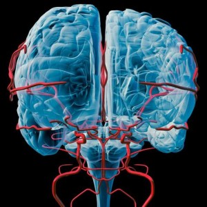
WELLNESS: Foods and activities that can reduce the risk for Alzheimer's disease Foods: Extra-virgin olive oil, coconut oil, most berries, cherries, elderberry, grape or grape seed, pomegranate, strawberry, leafy greens and cruciferous vegetables, rosemary, lemon balm, heperzine A, Panax ginseng, ashwagandha, ginkgo biloba, curcumin, coffee/caffeine, green tea, cinnamon, and dark chocolate. Supplements: alpha lipoic acid, acetyl l-carnitine, vitamin D, C and E, DHA, vinpocetin, Pyrroloquinoline quinone (PQQ), phosphatidylserine, magnesium, B vitamins (B12 and B6), niacin, co-Q10, N-acetyl cysteine,Luteolin, wild green oat extract,Nicotinamide riboside ,Bacopamonnieri – (Brahmi) , l-lysine (for the herpes virus), glutathione, and SOD (superoxide dismutase). Vega testing is an excellent way to determine which of these products your body might need. Nobody needs all of these. Also non-GMO foods and organic foods would be smart if you're dealing with advanced memory issues. The use of Stevia over artificial sweeteners would be recommended and artificial food coloring and other food additives could be an issue. Eat foods in their natural state. Activities that help: Exercise, learn something new, avoid toxic chemicals, toxic metals, PCBs, deal with stress, deal with depression, avoid processed foods and alcohol, the Mediterranean diet, the ketogenic diet, calories restricted diet, the Paleo diet, In a UCLA study, patients made dramatic lifestyle changes. They avoided simple carbs, gluten and processed foods. They increased their fish intake, took yoga and meditated. They were instructed to take melatonin, get adequate sleep, incorporate vitamin B-12, vitamin D-3 and fish oil. Within six months, nine of the 10 memory-loss patients saw a noticeable improvement in memory. One patient, who was in the late stages of Alzheimer's, did not show improvement. An ounce of prevention is worth a pound of cure.

CONDITION OF THE WEEK: LONGEVITY, DNA REPAIR AND CHIROPRACTIC DNA is the molecule of life. It contains our genes which make us what we are. DNA makes up our chromosomes which are held in the nucleus of our cells. DNA is very fragile and susceptible to damage from environmental stresses. It is estimated that our cells experience anywhere from 1,000 to 1,000,000 incidences of DNA damage PER DAY. If this DNA damage is left and not repaired, our DNA sequences cannot be read or will not be read properly. This will result in abnormal cell function or expression. Three things can then occur; senescence, which means the cells stop dividing, cell death and/or tumor growth. All three are not good things as they lead to poor health and premature aging. The good news is that our cells are equipped with numerous enzymes (too many to list) whose job it is to continually repair this DNA damage. This DNA repair can be measured through blood tests that measure serum Thiol levels. The serum Thiol level is an indicator of the level of DNA repair. The higher serum Thiol, the more DNA repair and the better your health and aging. In a landmark study published in the Journal of Vertebral Subluxation Research, chiropractors collaborating with researchers at the University of Lund found that chiropractic care could influence basic physiological processes affecting oxidative stress and DNA repair. These findings offer a scientific explanation for the positive health benefits reported by patients receiving chiropractic care. Long-term chiropractic care of two or more years was shown to re-establish a normal physiological state independent of age, sex, or nutritional supplementation. Symptom-free or primary wellness subjects under chiropractic care demonstrated higher mean serum thiol levels than patients with active disease, and produced some values that were higher than normal wellness values in non-chiropractic subjects. Wellness care pays big benefits
FUNNY BONES: I just read a book about Helium. It was so good that I can't put it down.@@ How is Christmas like your job? You do all the work and the fat guy in the suit gets all the credit.@@ Q: Why can't you trust an atom? A: Because they make up everything.@@ When Magnesium and Oxygen started dating I was like, "O MG!"@@ Dad: "Can I see your report card, son?" Son: "I don't have it." Dad: "Why?" Son: "I gave it to my friend. He wanted to scare his parents."


Courtesy of:
John H. Keefe III, D.C.
(918) 663-1111

IN THE NEWS:. Our body clocks cause wounds sustained at night to heal more slowly November 21, 2017 A new study has discovered how our body clocks cause wounds, such as cuts and burns, to heal approximately 60% faster if the injury happens during the day rather than at night. The study, published in Science Translational Medicine, is the first to show how our internal body clocks regulate wound healing by skin cells and optimize healing during the day. Our body clocks – or circadian rhythm – regulate nearly every cell in the human body, driving 24-hour cycles in many processes such as sleeping, hormone secretion and metabolism. Tests using skin cells – fibroblasts and keratinocytes – and also in mice showed that during the internal body clock’s ‘daytime’, wounds to the skin healed almost twice as efficiently as wounds incurred during the night. The researchers found that this effect was mirrored in humans with burns, by collaborating with clinicians and analyzing the records of 118 patients with burn injuries. The researchers found a key reason for this faster healing was that skin cells moved to the site of the wound to repair it much faster during the body clock’s daytime. “By taking these into account, not only could novel drug targets be identified, but also the effectiveness of established therapies might be increased through changing what time of day they are given.”
WELLNESS: Saccharomyces boulardii, or S. boulardii, is one type of probiotic out there. But there’s one major difference: it's not a type of bacteria. It's a yeast that happens to function like a probiotic in the body. For the past 30 or so years, doctors have recommended it to help patients with diarrhea. It helps to regulate the intestines and protect them from pathogens, including Clostridium difficile (c-diff), and other things that can damage the intestinal lining. It also modulates different parts of your immune system and keeps the intestinal barrier function up and running. Doctors have associated defects in the intestinal barrier function with different gastrointestinal diseases. S. boulardii can also help treat other ailments, such as lactose intolerance, and might even have a future in the treatment of acne, vaginal yeast infections, high cholesterol, fever blisters, and canker sores. It seems to improve the quality of life of people with irritable bowel syndrome (IBS), as well as Crohn's disease. Some striking effects of the probiotic were seen. Within weeks, obese and type 2 diabetic mice given S. boulardii exhibited reduced fatty liver, a 10% reduction in fat mass and body weight, and a 40 to 50% reduction in inflammatory markers. Furthermore, these improvements correlated with improvements to gastrointestinal tissue and several important changes in gut bacterial balance. This remarkable study provides new insight into the effects of S. boulardii and further supports the idea that probiotics could help reduce the passage of inflammatory molecules in your gut (known and endotoxin) into you blood, causing inflammation, disrupting your metabolism and damaging your health.

CHIROPRACTIC: Chiropractic Can Help Asthma, COPD and More…A Study Shows Regular Chiropractic Visits Can Help! One study has discovered that another way to keep your lungs breathing easier is with regular chiropractic visits. In September of 2016, the Journal of Physical Therapy Science published a study involving 30 subjects between the ages of 20 and 38 who were not currently being treated for any type of respiratory issue, nor were they experiencing any pain in their thoracic region. One-half were randomly assigned to an experimental group, which is the group that received actual spinal manipulation therapy. The remainders were assigned to the control, receiving sham treatments instead. Findings: These findings suggests that chiropractic care likely plays a more important role in healthy lung function than most people realize. This information may be helpful to patients who are already experiencing chronic respiratory issues and looking for relief, but it may also work to reduce the likelihood of lung-related diseases in the first place. As the COPD Foundation states, generally speaking, “once lung function is gone, it is gone for good.” That’s why they recommend engaging in activities which can maximize lung capacity. These include: getting some type of regular exercise, performing physically demanding exercises during the times when it’s easier to breathe, and staying indoors when extreme temperatures are expected or pollution is high.
FUNNY BONE: To avoid taking down my Christmas lights, I’m turning my house into an Italian restaurant.@@ Halloween is the beginning of the holiday shopping season. That’s for women. The beginning of the holiday shopping season for men is Christmas Eve.—David Letterman@@ What do Black Friday shoppers and the Thanksgiving turkey have in common? They know what it’s like to be stuffed and then jammed into a small place.@@ A mother gave her grown son two sweaters for Hanukkah. The next time he visited, he made sure to wear one. As he entered her home, instead of the expected smile, she frowned. “What’s the matter?” she asked. “You didn’t like the other one?”@@ Did you hear about the 2 silk worms in a race? It ended in a tie!@@Spraying graffiti inside an elevator is wrong on so many levels.
Visit our web site: keefeclinic.com-faceebook/keefeclinic.com
Courtesy of:
John H. Keefe III, D.C.
(918) 663-1111
IN THE NEWS: Common Acid Reflux Medications Promote Chronic Liver Disease Approximately 10 percent of the general population take a proton pump inhibitor (PPI) drug to block stomach acid secretions and relieve symptoms of frequent heartburn, acid reflux and gastroesophageal reflux disease. That percentage can be as much as seven times higher for people with chronic liver disease. Researchers at University of California San Diego School of Medicine have discovered evidence in mice and humans that stomach (gastric) acid suppression alters specific gut bacteria in a way that promotes liver injury and progression of three types of chronic liver disease. The study is published October 10 in Nature Communications. "Our stomachs produce gastric acid to kill ingested microbes, and taking a medication to suppress gastric acid secretion can change the composition of the gut microbiome," said senior author Bernd Schnabl, MD, associate professor of gastroenterology at UC San Diego School of Medicine. "Since we found previously that the gut microbiome — the communities of bacteria and other microbes living there — can influence liver disease risk, we wondered what effect gastric acid suppression might have on the progression of chronic liver disease. We found that the absence of gastric acid promotes growth of Enterococcus bacteria in the intestines and translocation to the liver, where they exacerbate inflammation and worsen chronic liver disease." There are inexpensive and readily available alternatives to PPIs. However, even non-PPI-based antacids (e.g., Pepto-Bismol, Tums, or H2 blockers such as Tagamet and Zantac) still suppress gastric acid to a lesser degree. While these other types of antacids were not tested in this study, Schnabl said any medication that suppresses gastric acid effectively could cause changes in gut bacteria and thus potentially affect the progression of chronic liver disease. Alternatively, non-pharmacological methods for managing heartburn are an option for some patients, including losing weight and reducing intake of alcohol, caffeine, and fatty and spicy foods. An FDA report cautions against high doses or prolonged use of PPIs, because they’ve been shown to increase the risk of infection, bone fractures and dementia. But the danger doesn’t stop there. All acid stopping drugs (not just PPIs) inhibit nutrient absorption, promote bacterial overgrowth, reduce resistance to infection and increase the risk of cancer and other serious diseases.
CHIROPRACTIC: American Chiropractic Association (ACA) has announced a national health care observance
October is National Chiropractic Month
DO YOU KNOW SOMEONE WITH A HEALTH PROBLEM OR IN PAIN?
INVITE YOUR FAMILY OR FRIENDS TO THE PARTY Who do you know who is hurting? Who do you know that you have been trying to get started at Keefe Clinic?
Now is the time to get your spouse or child started on the road to good health.
Complimentary DIAGNOSTIC WORK UP DURING OCTOBER
Complimentary initial exam and one X-ray.
Tell someone today
WELLNESS: Best foods for good digestion Yogurt-has bacteria that is essentially good for your gut. You need to check their labels for "live and active culture" to reap the digestive benefits out of it. Banana-are very effective in treating gastric problems as they are helpful in restoring bowel function and can help treat diarrhea. They are rich in electrolytes and potassium which help in restoring good digestive health. Ginger-is a spice that has many benefits for digestive health. It can help cure motion sickness, nausea, vomiting, gas and loss of appetite. The ideal consumption would be 2 to 3 g every day. If you have more than that, it may cause heartburn. Beet root-are very good source of fiber, potassium and magnesium. They are an excellent cure for problems like constipation. Apple-are also rich in bacteria that are helpful in maintaining a good gut health. Avocado-are among the best sources of fiber and fruits. This helps in maintaining a mucosal lining in the gastrointestinal tract, which helps in the digestive process. Cod liver oil-source of the vitamin A and C, cod liver oil helps in maintaining a good digestive health. Blueberry-a very good source of fiber and vitamin C. They are loaded with cancer fighting properties. Kiwi-is loaded with minerals and nutrients which are very good for your gut health. It contains vitamin C and E, linolenic acid, magnesium, potassium, actinidin, fatty acids and pepsin which are good for your digestive health. Papaya-rich in papain which helps break down proteins in your stomach. Papaya also has anti-inflammatory properties which can help and soothing the stomach. It also helps relieve problems like food allergies and heartburn.
FUNNY BONE: TEACHER: Glenn, how do you spell crocodile? GLENN: K-R-O-K-O-D-I-a-L TEACHER: no, that’s wrong. GLENN: maybe it is wrong, but you asked me how I spell it.@@ I went to buy some camouflage trousers the other day but I couldn't find any.@@ If you jog in a jogging suit, lounge and a lounging pajamas, and smoke in a smoking jacket, why would anybody want to wear a windbreaker?@@ A police car pulled me over near the high school where I teach. As the officer asked me for my license and registration, my students began to drive past. Some honked their horns, others hooted, still others stopped to admonish me for speeding. Finally the officer asked me if I was a teacher at the school, and I told him I was. I think you’ve paid your debt to society, he said with a smile, and left out given me a ticket.
Visit our web sites: keefeclinic.com & facebook.com/keefeclinic
Courtesy of:
John H. Keefe III, D.C.
(918) 663-1111
American Chiropractic Association (ACA) has announced a national health care observance
October is
National Chiropractic Month
DO YOU KNOW SOMEONE WITH A HEALTH PROBLEM OR IN PAIN?
INVITE YOUR FAMILY OR FRIENDS TO THE PARTY
Who do you know who is hurting? Who do you know that you have been trying to get started at Keefe Clinic?
Now is the time to get your spouse or child started on the road to good health.
Complimentary
DIAGNOSTIC WORK UP DURING OCTOBER
Complimentary initial exam and one X-ray.
Tell someone today

IN THE NEWS: Do germs trigger type 1 diabetes? Germs could play a role in the development of type 1 diabetes by triggering the body’s immune system to destroy the cells that produce insulin, new research suggests. Scientists have previously shown that killer T-cells, a type of white blood cell that normally protects us from germs, play a major part in type 1 diabetes by destroying insulin producing cells, known as beta cells. Unlike type 2 diabetes, type 1 diabetes is prevalent in children and young adults, and is not connected with diet. In previous studies the Cardiff team isolated a killer T-cell from a patient with type 1 diabetes to view the unique interaction which kills the insulin-producing beta cells in the pancreas. “In this new study, we wanted to find out what was causing these T-cells to kill beta cells. We identified part of a bug that turns on killer T-cells so they latch onto beta cells.” The research, published in The Journal of Clinical Investigation, provides a first ever glimpse of how germs might trigger killer T-cells to cause type 1 diabetes, but also points towards a more general mechanism for the cause of other autoimmune diseases. NOTE: Probiotics/friendly bacteria help keep the population of bacteria in check and balanced. Get a daily supply of friendly bacteria. Introduce your kids to Kefir or other cultured foods.

WELLNESS: Apricot seeds, B-17/Laetrile Apricot seeds are similar to other nuts - rich in healthy fats and a source of fiber and iron. There are two types of apricots seeds, or kernels. Sweet apricot seeds are sometimes sold as snacks or for baking, and they contain very little or no amygdalin. Bitter apricot seeds, on the other hand, do contain amygdalin. Laetrile is one of the most popular and effective alternative cancer treatments available. In order to increase the efficiency of Laetrile, it is recommended to follow a strict nutrition diet and also purchase other numerous supplements. Laetrile targets and kills cancerous cells, while repairing the immune system to fend off future possibilities of cancer. B-17 helps boost immunity, relieves pain, lowers high blood pressure, relieves constipation, can help with respiratory disease, has anti-inflammatory properties, protect cells from damage, antimicrobial properties, helps maintain proper pH balance. Recommended usage is 2-3 daily. However well-known authors like Marcus D. Adams claims that you can have up to 24 to 40 kernels a day spread throughout the day. You have to start with 2 and see how much your body can take on a daily basis.
FUNNY BONE: If I won the award for laziness, I would send somebody to pick it up for me.-@@ Don't know where your kids are in the house? Turn off the internet and they'll show up quickly.@@ As your best friend I'll always pick you up when you fall, after I finish laughing.@@ When you wake up at 6 in the morning, you close your eyes for 5 minutes and it's already 6:45. When you're at work and it's 2:30, you close your eyes for 5 minutes and it's 2:31.-@@ Scene: A radio newsroom. Caller: I just wanted to let you know you’re off the air. Host: Yes, we know. The engineers are working on it. Caller: It would be nice if you put something on the air that says that.@@ The food at the sandwich shop I frequent is good, but any deviation from the norm throws the staff. I once told a clerk that I wanted only half a sandwich. His reply: “What am I going to do with the other half?” A week later, when I told another clerk the same thing, she responded, “Do you want the top or the bottom?” Carole Holder, Norman, Oklahoma
Visit our web sites: keefeclinic.com & facebook.com/keefeclinic
Have you been told ……
“There is no cure for what you have, no one can help you”.....
Really?
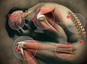
Many patients have been told this by their medical doctor and yet have gone on to find help through natural healthcare and chiropractic. It's not that the medical doctors were lying to them but what they were really saying is drugs and surgery can't help you. Their language is unfortunate because that traps many people with conditions they live with the rest of their lives. It's also sad that medical doctors are brainwashed in school into believing that if drugs and surgery can't help the patient in front of them, nothing else will.
Chiropractic and natural healthcare approaches the body with completely different eyes. We might do some of the same tests like lab work, MRIs or x-rays and physical exams but we interpret the information completely different in some cases. The focus of drug therapy is to find some chemical that will suppress your symptoms. It's like with type II diabetes where the body is not responding to the insulin it's producing so they use chemicals to force the sugar out through the kidneys. This doesn't help the cells that need sugar for proper metabolism to get the sugar they need it just limits the danger of ever increasing blood sugar levels. The natural approach focuses on nutritional and lifestyle changes that help the cells recognize and respond properly to the insulin.

It's been said there are no incurable diseases just some incurable patients. The process of getting well can be complex and difficult in some cases the motivation to make the changes in lifestyle and diet are more than some people are willing to do thus they carry their condition to an early grave. The key in any health problem is breaking the code for that disease and to do that sometimes it's as simple as doing basic blood work and understanding the nutritional components of those findings but in some cases it's more complicated, requiring innovative tools to break the code. That's one reason we use a muscle reflex testing protocol that we call Vega testing. The founder of Vega testing was a German medical doctor/dentist who developed this test that included both the protocol of testing along with a sophisticated Ohm's machine to study the response from the body. Because that machine was both expensive and subject to operator error we decided to use a muscle reflex test to work the protocol, it is more objective and quicker. In Vega testing we monitor muscle reflexes in response to certain diagnostic ampules to help us break the code of the disease, understand the underlying components. We can quickly, in a lot of cases, find the underlying toxicity, emotional component, nutritional imbalance that’s allowing the condition to remain in the body. Your body has an ability to heal itself but that ability can be interfered with because of different blockages. Our goal is to determine what those blockages are and remove them allowing your body what it does best, heal.
The basic approach that we use at Keefe Clinic includes determining what body type you are so we can establish the right ratio of fats, carbs and proteins for your particular genetic makeup. Along with that diet, detoxification, possibly an anti-inflammatory diet, and neurological balancing and establishing the necessary neurological pathways so the body can regulate itself and in turn heal itself. Targeted nutritional therapy, exercise, stress handling are just some of the techniques that were used to help the body.
LINK: Vega testing
Health is a choice

Working with patients for over 42 years I’ve learned the importance of proper lifestyle choices. It’s been said there are no incurable diseases just some incurable patients. Outside of rare genetic disorders most people have the inborn ability to live a healthy, pain-free life. But that ability requires making quality life choices. I’m pretty sure most people realize what shape your house would be in you never cleaned it in 20 years. A similar thing is true about your body; if you’re not proactive in taking care of your health then complicated health issues can develop that take time to repair. Unfortunately, in most cases, we are unaware that our bodies are breaking down because we have no obvious symptoms. It’s similar to drinking water, as we age our sense of thirst diminishes and we can be significantly dehydrated and not even know it.
A lot of times it is just simple things that need to be addressed in order to maintain excellent health. Movement is important, you don’t have to be conditioning for the Olympics to stay in good health even three days a week of walking or other exercise can have a significant benefit. Rest and relaxation including refreshing sleep are crucial on regulating stress hormones that can have a profound effect on your health over time. Positive mental and spiritual attitude have a healing effect on the mind as well as the body, practicing an act of kindness not only helps others in our society but floods your body with important hormones and chemicals that not only give you a sense of well-being but have health promoting benefits. Since the nervous system controls and coordinates everything in the body maintaining a stress free nervous system through regular chiropractic adjustments are crucial. Chiropractic keeps the communication channels open and allows the body to self-regulate and heal. And what you put in your mouth is crucial, it has been said that, “We dig our own graves with a fork.”
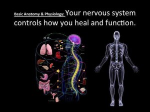
At Keefe clinic we try to engage each of these five laws of health to help you attain and maintain good health. No matter what pharmaceutical companies tell you drugs can’t heal you they can only manipulate your perception of symptoms. We not only want to encourage you to make healthy choices but also to provide tools to help you maintain the best health possible. With Vega testing we not only pick up problems that are developing but zero in on the best product or formula to restore normal function.
Since inflammation is a major destroyer of health in our society eating anti-inflammatory foods are essential, fish oils are probably the most important way to control inflammation.
CLICK TO ORDER
EPA DHA 300 is an excellent mercury free fish oil to help you maintain better health, 2 to 3 capsules a day will not only keep your heart healthy but help reduce inflammation in your body that can lead to arthritis and other health issues.
Antioxidants are another important addition to controlling chronic disease.

CLICK TO ORDER
Pomegranate plus is a blend of the most potent antioxidant foods and taking one or two a day is an excellent way to support your health. Remember with testing we can be more specific with what your body needs.
To your better health,
Dr. Keefe
If you have not set up your account at natural partners then go to npscript.com/Keefeclinic and set up an account (if a code is asked for use 6631111). There is no charge to set up this account and they do not sell email addresses. Once the account is set up you can order any of their over 9000 products in the comfort of your own home and have them shipped to your door. Note: for period of time you can get free shipping, order today! For help ordering call 888-950-2767
Courtesy of:
John H. Keefe III, D.C.
(918) 663-1111

IN THE NEWS: ‘What the Health’ — Where This Vegan PR Film Went Wrong The documentary, “What the Health,” ignores accumulated evidence against sugar in a misguided effort to promote vegan ideology. According to the film, the focus on sugar as a contributor to obesity, diabetes and ill health has steered people away from the real culprits, which they claim are meat and animal fat. For example, Dr. Neal Barnard, adjunct associate professor of medicine at the George Washington University School of Medicine and Health Sciences and president of the Physicians Committee for Responsible Medicine, claims that diabetes is not caused by a high-carb, high-sugar diet. In his view, diabetes is caused by fat buildup caused by a meat-based diet. To treat diabetes, Barnard recommends a low-fat vegetarian diet, free of any and all animal products, without any restrictions on carbohydrates. The sugar industry promotes the myth that saturated fat is to blame for weight gain and ill health, not sugar, along with the thoroughly debunked energy balance theory. Fortunately, some great books have now been written exposing the history and extent of the cover-ups. Two examples are science journalist Gary Taubes' book, "The Case Against Sugar," and Marion Nestle's "Soda Politics: Taking on Big Soda (and Winning)." While a high-vegetable diet is certainly beneficial, the low-fat, unrestricted-carb recommendations are not. Low-carb, high-fat diets have proven superior for controlling insulin resistance, which is the hallmark of obesity and metabolic dysfunction. NOTE: This article points out the fight within nutritional science between the high protein low carb diet and the high complex carb low-fat diet. The truth is both diets are correct for different populations, 60 to 70% of the population should be on a high protein low carbohydrate diet where 30 to 40% of the population need to be on a low fat high complex carb diet. It’s a matter of genetics, we are not all the same. KNOW your body type!

WELLNESS: Are you getting enough fiber? One of the most underserved nutrients today is fiber. Typically missing or very low in packaged and fast foods, fiber intake for most Americans is woefully low. Adults need between 20 to 30 grams of fiber every day for optimal health, but most Americans get only about 15. I believe the ideal is closer to 32 grams. What potential benefits might you expect when you increase the amount of fiber you eat each day? Fiber: Assists in managing your weight by helping you feel full longer Helps promote regularity Helps support your heart health Helps support healthy GI function. Your body needs two different types of fiber: soluble and insoluble. Soluble fiber helps slow down your digestion and helps maintain glucose and blood cholesterol levels already in the normal range. Good food sources are nuts, seeds, apples, and blueberries. Insoluble fiber helps propel food through your digestive tract and promotes regularity. Good food sources include dark green leafy vegetables, green beans, celery, carrots, and brown rice. Many whole foods, especially fruits and vegetables, contain both soluble and insoluble fiber, a real nutritional bonus.

CHIROPRACTIC: How Does Chiropractic Work? Many people seek chiropractic care when their back goes out or their neck tightens up. But how does this form of care actually work? What are the benefits of receiving chiropractic care for nerve dysfunction compared with other healthcare options? Let’s take a look! First, let’s discuss how the nervous system “works.” We have three divisions of the nervous system: the central, peripheral, and autonomic nervous systems. The central nervous system (CNS) includes the brain and spinal cord, and it’s essentially the main processing portion of the nervous system. The spinal cord is like a multi-lane highway that brings information to the brain for processing (sensory division) and returns information back to the toes, feet, legs, and upper extremities from which the information originated (motor division). For example, hiking on a mountain trail or simply walking requires constant input to and from the CNS so we can adjust our balance accordingly and not fall. These “sensory-motor pathways” are essential and allow us to complete our daily tasks in an efficient, safe manner as information is constantly bouncing back and forth between the brain and the rest of the body. The peripheral nervous system (PNS) includes a similar sensory/motor “two-way street” system relaying information back and forth from our toes/feet/legs and fingers/hands/arms to the spinal cord (CNS). And if this isn’t complicated enough, we also have “reflexes” that, for example, allow us to QUICKLY pull our hand away from a hot stove to minimize burning our fingers. Reflexes allow the information to “skip” the brain’s processing part so quicker reactions can occur. The autonomic nervous system (ANS) includes the sympathetic and parasympathetic divisions that basically “run” our automatic (organ) functions like breathing, heart rate, digestion, hormonal output, and more. There is constant communication between the ANS, PNS, and CNS that allow us to function in a normal, balanced way… unless something disrupts them. There are obvious conditions that interfere with this communication process that include (but are not limited to) diabetes (with neuropathy), frost bitten or burned fingers, peripheral nerve damage from conditions like carpal/cubital tunnel syndromes, thoracic outlet syndrome, and/or pinched nerves in the neck, mid-back, low-back spinal regions, as well as conditions such as multiple sclerosis (MS), Guillain-Barre Syndrome, after a stroke (spinal cord or brain), and after trauma with resulting fractures where nerve, spinal cord, and/or brain damage can occur. These are “obvious” reasons for delayed or blocked neurotransmission. There are many other less obvious injuries or conditions that can result in faulty neuromotor patterns and nerve transmission of which chiropractic services can benefit. The “subluxation complex” is a term some chiropractors use to describe the compromised nerve transmission that may occur if a nerve is compressed or irritated due to faulty bone or joint position along the nerve’s course. Reducing such nerve compression typically allows for a restoration of function. A good illustration of this is a patient who suffers from a herniated disk in the neck with numbness and tingling down the arm to the hand. The goal of treatment (for all healthcare professionals) is to remove the pinch of the nerve. To realize this goal, doctors of chiropractic utilize spinal manipulation and mobilization in addition to other non-surgical, non-drug approaches that may include exercises, nutritional advice, home-care such as a cervical traction unit, and other anti-inflammatory measures (ice, modalities like low level and class IV laser, electric stimulation, pulsed magnetic field, and more). Given the minimal side-effect risks and well-reported benefits, it only makes sense to try chiropractic FIRST and if you’re not satisfied, your doctor will help you find the next level of care.
FUNNY BONE: Ah, marriage. I was standing in front of the bathroom mirror one evening admiring my reflection, when I posed this question to my wife of 30 years: “Will you still love me when I’m old, fat, and balding?” She answered, “I do.”@@ As the music swelled during a recent wedding reception, my hopelessly romantic husband squeezed my hand, leaned in, and said, “You are better looking than half the women here.”
Visit our web site: keefeclinic.com or faceebook/keefeclinic.com

Proper Health starts with the correct acid-alkaline balance in your body. The pH level (acid - alkaline measurement) of our internal fluids affects every cell in our bodies. Extended acid imbalances of any kind can overwhelm your body, and lead to health complications.
Just as the body regulates its temperature in a rigid manner, so will it manage to preserve a very narrow pH range - especially in the blood. As a matter of fact, the body will go to such great lengths to maintain a blood pH of 7.365 that it will even create stress on other tissues, body systems, and organs to do so. Chronic acidity will interrupt all cellular activities and functions - it interferes with life itself.
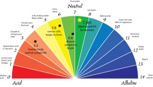
CLICK HERE
When the pH of the body gets out of balance (too acidic), we may experience low energy, fatigue, excess weight, poor digestion, aches and pains, and even more serious disorders.
The cycle of acidity begins primarily as a result of three things:
1. Ingesting acids. Eating too many acidifying foods like processed sugar, meats, dairy, coffee, alcohol, etc. Create an acid ash in the body. These acids can overload the body's ability to neutralize them.
2. Creation of acids. Pathogens and microforms create acidifying toxins in the body. As the body becomes more and more acidic, bad bacteria, yeasts and other microforms proliferate in the body. Since these organisms are living, they eat as well as create resulting toxins. These toxins are often very acidifying.
3. Improper elimination of acids. Not all acids are the same; some are weak and some are strong. Weak acids like citric acid are much easier to neutralize than strong acids like uric acid. The body uses many systems in order to buffer acids including breath, mineral reserves, and fat. When the body's buffering systems become compromised, excess acids build up.

CLICK HERE
The process of reestablishing acid-alkaline balance begins with proper diet and nutrition. This includes eating alkalizing foods (vegetables, low sugar fruits, etc.), super hydration (drinking plenty of alkaline water), and proper supplementation.
Keeping yourself BALANCED is your first line of defense in fighting any disease and alkaline water creates a major impact in balancing the body's pH.
CLICK TO FIX
A body that is too acidic provides the ideal environment for diseases to manifest and thrive in. For example, accumulated acid waste in the joints encourages inflammation and arthritis. It turns acidic wastes into solids and stores them in fatty tissues resulting in hardening of the arteries and excess weight. Our body also tries to buffer the effect of this excess acid by taking calcium from the bones, and magnesium needed for heart health.
Therefore, one of the keys to great health is keeping the body pH properly balanced. Because of the predominance of hydroxyl ions produced, making the water highly alkaline, drinking plenty of alkaline water will help dissolves accumulated acid waste and will balance your body pH.
Restoring the pH balance in the body.

CLICK TO ORDER
Alkaline water can neutralize the acidity of the body caused by stress, modern diet, air pollution, and many bottled waters.
- A higher pH in the body reduces the need for fat and cholesterol to protect the body from damaging acids.
- Alkaline water is negatively charged and an "antioxidant." Antioxidants reduce cellular and DNA damage caused by free radicals. Negatively charged alkaline water creates energy by giving up ions to positive ions.
- Alkaline water tastes lighter with a pleasantly sweet flavor.
- Improves the body's absorption of essential nutrients
- Improves body function by cleaning your cells from the inside out.
- Using water with a higher pH level improves the taste of beverages and food.
- Improves your immune system function to help you fight diseases.
- Cooking with alkaline water improves the taste and quality of foods and using acidic water when boiling eggs improves their quality
Understanding pH Values
For those not familiar with pH scales:
Pure acid is pH 0
Pure alkaline is pH 14
Neutral (neither acid nor alkaline) is pH 7
pH 7 is ten times more alkaline than pH 6
pH 8 is 100 times more alkaline than pH 6
pH 9 is 1,000 times ore alkaline than pH 6
How much do I drink?

CLICK FOR INFO
Your water needs are based on your body weight. Half your body weight is your cleansing dose and 80% of that is your minimum because otherwise you're dehydrated.
100 pound person, 50 ounces is their cleansing dose and 40 ounces is a minimum of fluids they should drink every day.
 How much alkaline water should I drink? That depends on your pH. We carry very sensitive pH paper for determining your urine pH which is the pH we base your need for alkaline water on. If you're a vegetarian and your pH is already alkaline then you can just drink regular filtered water. But if your pH is lower than 6.4 then you need to drink alkaline water. I would recommend 1 L a day minimum. Depending on your weight more might be indicated.
How alkaline of water should I drink? Water can come from about 7.8 to 10. You can drink water that is to alkaline for you and it is best to be tested at our office to determine the best pH. Waters with a pH of 9 to 9.5 tend to test the best, but your situation might be different.

CLICK TO ORDER
Both saliva and urine pH should be 6.4. When you maintain that then your blood and cells will be able to maintain the pH that is right for them. Saliva pH indicate your digestive pH whereas urine pH indicates your body pH. So for determining your need for alkaline water urine pH is the one to watch. Also it is recommended not to drink alkaline water with meals. If you're going to drink water with a meal I recommend squeezing several lemon or lime slices in the water to make it acidic. Lemon and lime will be alkaline on your body yet acid in your stomach.

I don't keep up with drug sales let alone the actions and side effects of all the latest drugs. But I would think antacids must be making pharmaceutical companies literally billions of dollars each year. (The January 8, 2001 issue of Business Week mentions that Priolsec is the top selling prescription drug in the world. It earns Astra Zeneca (the drug’s maker) 6 billion dollars per year.)
CLICK FOR INFO
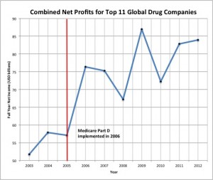
CLICK FOR MORE
One of the reasons that antacids sell so well is that they stop the pain in 60-70% of the cases. (A recent online survey of over 4200 patients taking Priolosec.) If you're troubled with chronic acid reflux and you can take a pill and all the pain just disappears, it seems like you're doing the right thing, doesn't it? Before you answer that let me explain the digestive process and then ask you that question again.
Digestion is literally a miracle. All the different types of foods and yet your body knows the exact enzymes and the correct amount to properly breakdown each food to its simplest component. Complex carbohydrates are broken down into simple sugars, proteins are broken down into their individual amino acids and fats and oils are reduced to their individual fatty acids. And then the process of properly absorbing these nutrients along with the vitamins, minerals and enzymes plus other things that were still learning about. If you eat different foods you get different kinds and amounts of enzymes to digest them.
CLICK FOR MORE

CLICK HERE
As you chew your food you help break it down for your stomach as well as blend it with saliva which helps start the whole process. As the food enters the stomach it starts to churn and release digestive enzymes. Hydrocloric acid (HCl) is one of the most important enzymes that the stomach releases. The amount of HCl that leaves the stomach with the food entering the small intestines stimulate fluids from the liver and gallbladder as well as enzymes from the pancreas to complete the digestive process. It is essential that the right amount of HCl enters the small intestines to begin a wavelike motion called peristalsis that help move the food throughout the intestines. If the level of HCl is less than ideal then every other step in the digestive process is compromised.

CLICK HERE
It's like when you have a row of dominos stacked together and push the first one over a chain reaction happens. Without HCl the rest of the digestive process is severely compromised. If you don't produce the right amount of enzymes then you cannot breakdown your foods properly. If you don’t break your foods down properly you can not absorbe nutrients that your body requires. Studies of individuals who have used antacids over a long peiord of time find evidence of malnutrition. Doesn't that make sense? What do antacids do? They severely reduced if not completely block the release of HCl. Remember the chain of dominos.

CLICK HERE
When you consistently reduced the amount of HCl that is released from your stomach that causes a series of changes in the lining of your stomach. A healthy stomach has a mucous lining that protects the stomach from its own acids. When the body senses that this coating has been reduced it automatically reduces the amount of HCl it secretes. As this happens the stomach will churn more to help break down the foods. After a series of processes, this increase of abnormal stomach contractions can force what little bit of HCl you produce up into the esophagus.

CLICK HERE
This can cause a burning or painful sensation from your stomach up into the back of your throat. This is called acid reflux. This condition is rarely due to the overproduction of HCl. If this problem is not corrected over a lifetime the chronic inflammation to the cells can lead to cancer. But be forewarned, the chronic suppression of HCl with antacids will lead to incompletely digested food irritating your intestines and increasing your chance for colon cancer. Usually the mechanism I described above is leading to the inappropriate release of HCl into the esophagus. It is true eliminating the amount of HCl you produce will relieve symptoms. It's also true that these drugs will relieve you of the normal digestive process.
CLICK HERE
Now the main question is this- is the choice between symptom relieve with drugs or suffering pain with each meal? No! By properly correcting the dysfunction within the digestive system you can, not only restore proper function without drugs, you can also overcome your acid reflux.
CLICK HERE
There are several principles to good digestion. Completely chewing your food, not combining proteins with sweet fruits, don't eat too quickly, eat foods that are consistent with your body and blood type, too much dairy with a meal could interfere with the digestive process and watch the junk food. Fiber helps move the food through your digestive system.
CLICK HERE
A lot of time patients with digestive problems also notice that they have discomfort between the shoulder blades. Neurologically this is very significant. The nerves between the fourth thoracic and eight thoracic are some of the most important controlling nerves to your digestive system. If you carry nerve pressure in your upper back then your digestive system is most likely compromised. Now remember, you can have nerve pressure without back symptoms, though alot of patients with acid reflux know what I'm talking about when I talk about discomfort between their shoulder blades.

CLICK TO ORDER
B-vitamins are very important when it comes to digestion and because you can't store B-vitamins. A good percentage of patients with digestive problems also have deficiencies in some of their B-vitamins. We also use targeted nutritional formulas to help restore the normal lining of the stomach along with diet and Chiropractic adjustments.

CLICK HERE
The care I just outlined for acid reflux is also extremely beneficial from most other digestive problems. Chiropractic care is the most effective treatment on the planet for digestive disorders. Most other approaches, even with gastroenterologists, focus primarily on symptom relieve and not correction.

CLICK HERE
Now let me ask you that question again and tell me if you might have a different answer. If you're troubled with chronic acid reflux and you can take a pill and all the pain just disappears, it seems like you're doing the right thing, doesn't it? Are you?
Courtesy of:
John H. Keefe III, D.C.
(918) 663-1111

IN THE NEWS: Exposure to violence hinders short-term memory, cognitive control Being exposed to and actively remembering violent episodes—even those that happened up to a decade before—hinders short-term memory and cognitive control, according to a study published in the Proceedings of the National Academy of Sciences (PNAS). The study, which was co-authored by Princeton University's Pietro Ortoleva, examined more than 500 civilians in Colombia, a country that has experienced both urban violence and rural warfare within the past two decades. The findings demonstrate the long-lasting effects of violence on cognition and memory recall and highlight the need for policies that provide proper therapy for those coping with violence. "Memory and cognitive control impact how people do in school, how they perform at work and if they can keep their jobs, and how they fare in life in general, which all has significant impacts on the economy as a whole," said Ortoleva, a professor of economics and public affairs at Princeton's Woodrow Wilson School of Public and International Affairs. "Our results demonstrate that exposure to violence can have effects on cognitive functions," said Ortoleva. "Besides the obvious negative effects on the physical and psychological well-being, this may lead to vicious cycles: both poverty and violence hinder the ability to develop for a person or a group, which in turn may generate further poverty and violence. Emotional freedom technique is something that we use in our office to help the mind process emotional injury. If you feel that you are dealing with issues like this asked Dr. Keefe on your next visit for an evaluation.
WELLNESS: Heartburn Drugs Increase Mortality Proton pump inhibitors are commonly used to treat heartburn, but have been shown to do far more harm than good, raising your risk of kidney disease, dementia, heart attacks and premature death. Research clearly shows PPIs are outrageously overprescribed and misused, and do far more harm than good. There are many alternative treatment strategies that can help ease this painful problem without the serious side effects associated with PPIs, which include kidney disease, pneumonia, osteoporosis, hip fractures, dementia and an increased risk for heart disease, heart attacks and premature death. Most acid reflux cases are related to insufficient amounts of stomach acid, food allergies, Helicobacter pylori imbalance and/or hiatal hernia. Gallbladder disease can also be mistaken for heartburn. The long-term answer to heartburn and acid indigestion is to restore your natural gastric balance and function. The most important step is to eat real food, as processed foods and sugars are a surefire way to exacerbate acid reflux. Reseeding your gut with beneficial bacteria, either from traditionally fermented foods or a high-quality probiotic supplement, is also important. Aside from that, there are a number of natural substances that can help address the root of the problem. To address heartburn, eat unprocessed food and fermented foods, and take supplemental digestive enzymes. Foods such as papaya, pineapple and ginger can also help, as well as a number of other supplements.

CONDITION OF THE WEEK: Phantom organ symptoms A lot of people have heard of phantom limb syndrome where patients who have lost an arm or leg can still feel that appendage. The reason for that is the place in the brain where that limb is controlled and coordinated is still there even though the limb is gone. In a lot of cases the trauma that caused the limb to be lost produced subluxations that press on the nerves that used to go down the arm or leg. Another phantom pain is when an organ has been removed like a gallbladder or spleen from an accident. These patients can still suffer symptoms even though they no longer have the organ. Both chiropractic adjustments and even nutritional therapy can help these annoying complaints, they can be resolved in a high percentage of the cases. Chiropractic care and nutritional therapy should be your 1st choice of treatment. Chiropractic 1st, drug 2nd, surgery last.

FUNNY BONE: A Canadian psychologist is selling a video that teaches you how to test your dog’s IQ. Here’s how it works: If you spend $12.99 for the video, your dog is smarter than you.@@ I’d never had surgery, and I was nervous. “This is a very simple, noninvasive procedure,” the anesthesiologist reassured me. I felt better, until … “Heck,” he continued, “you have a better chance of dying from the anesthesia than the surgery itself.”@@ Aerial Photos--A photographer for a national magazine was assigned to take pictures of a great forest fire. He was advised that a small plane would be waiting to fly him over the fire. The photographer arrived at the airstrip just an hour before sundown. Sure enough, a small Cessna airplane was waiting. He jumped in with his equipment and shouted, "Let's go!" The tense man sitting in the pilot's seat swung the plane into the wind and soon they were in the air, though flying erratically. "Fly over the north side of the fire," said the photographer, "and make several low-level passes." "Why?" asked the nervous pilot. "Because I'm going to take pictures!" yelled the photographer. "I'm a photographer, and photographers take pictures." The pilot replied, "You mean you're not the flight instructor?"
Visit our web sites: keefeclinic.com & facebook.com/keefeclinic
Courtesy of:
John H. Keefe III, D.C.
(918) 663-1111

IN THE NEWS: Oxygen and hyperbaric oxygen therapy reverses brain damage in drowned toddler Medical researchers report the case of the reversal of brain volume loss in a 2-year-old drowning victim unresponsive to all stimuli treated with normobaric oxygen (oxygen at sea level) and hyperbaric oxygen therapy. The patient and family then traveled to New Orleans where 78 days post-drowning, Dr. Harch began treating her with hyperbaric oxygen therapy. She "dove" in a hyperbaric chamber for 45 minutes a day, five days a week for 40 sessions. At the beginning of each session, the patient showed visually apparent and/or physical examination-documented neurological improvement. After 10 HBOT sessions, the patient's mother reported that her daughter was "near normal" except for gross motor function, and physical therapy was re-instituted. After 39 HBOT sessions, the patient exhibited assisted gait, speech level greater than pre-drowning, near normal motor function, normal cognition, improvement on nearly all neurological exam abnormalities, discontinuance of all medications, as well as residual emotional, gait and temperament deficits. Gait improvement was documented immediately upon returning home. An MRI at 27 days following HBOT session 40 and 162 days post-drowning demonstrated mild residual injury and near-complete reversal of cortical and white matter atrophy.
WELLNESS: MUSIC THERAPY CLINIC at ORU The Music Therapy program at Oral Roberts University is built upon an integrated music therapy clinical program of Cognitive-Behavioral Music Therapy and Neurologic Music Therapy (NMT). The integrated program develops clinical knowledge and treatment based on analysis of individuals’ cognitive, speech & language, social/emotional (psychological), and physical behaviors, and prepares music therapy students to utilize scientific evidence regarding human perception and the production of music to treat disorders of the human nervous system. The Music Therapy program is dedicated to providing quality music therapy services to the local community through the operation of a professional Music Therapy Clinic. This clinic will allow students to gain professional experience evaluating and treating patients, while being supervised by three board-certified music therapists, Dr. Hayoung Lim, Dr. Hyun-Jung Lee, and Jacqueline Cox. In every music therapy treatment, music is used as a tool to reach nonmusical goals, whether physical, cognitive, emotional, social or spiritual. The techniques used during sessions are based on research and extended collegiate study. Among many possible goals addressed by music therapists might be: Increasing memory recall Improving motor coordination Increasing attention to task Improving steadiness of gait Improving speech and communication Increasing reality-based thinking Decreasing anxiety Decreasing pain Adjusting mood Developing bonds among family members Increasing coping skills
CHIROPRACTIC: Achy joints, migraines and stomach cramps? Ditch the painkillers and learn how to EAT your pain away 1. GET YOUR FIVE-A-DAY It is one health message that no-one can claim to be unaware of - eating five pieces of fresh fruit or vegetables each day is beneficial to a person's general health and wellbeing. However, a specific benefit of eating a rainbow of colours, is that it can also help to get rid of pain 2. THE 'F'-WORD Fats in our diet are metabolised in the body and turned into 'local hormones', known as prostaglandins. 'There is evidence that the omega-3 fatty acids reduce it. In contrast, omega-6 fatty acids are linked to increasing inflammation. 3. BUILDING BLOCKS Protein is essential for repair and healing in the body, but high levels of protein from meats may actually have the opposite effect. 4. SPICE IT UP! Include plenty of gentle spices in your meals.The spices turmeric, cayenne and ginger are known to have an anti-inflammatory action in the body. 5. HOLD ON TO YOUR WATER BOTTLE Water transports nutrients to where they are needed in the body and removes toxins, waste products and dead cells, which are produced in higher quantities when there is inflammation. REASONS TO DRINK FILTERED WATER More than half of the chemicals found in U.S. drinking water are unregulated, and many of the legal limits may be too lenient for safety. Firefighting foam used on military bases and other fire testing sites has contaminated many water tables across the U.S. with PFCs — chemicals linked to thyroid disease and cancer. Agricultural runoff, prescription drugs flushed down the sewer and aging water pipes is other significant sources of water pollution. Chlorine and fluorine are just some of the chemicals that can damage arteries and led to heart disease. Ask us about a filtration system. 6. SAY 'NO' TO... Sugary foods and refined, Coffee, Fizzy drinks, Saturated Fats.
FUNNY BONE: Judge: “Why did you steal the car?” Man: “I had to get to work.” Judge: “Why didn’t you take the bus?” Man: I don’t have a driver’s license for the bus.@@ I have a bumper sticker saying, "Honk if you think I'm sexy". Some days I just stand at a green light till I’m feeling good about myself.@@ Wife sends a text message to her husband on a really cold winter morning: Windows are totally frozen, will not open. Husband replies: “Carefully pour some warm water over it and tap the edges first with your hand, if that doesn’t work, then gently with a hammer.” 15 minutes later, the wife texts back: “Oh no, I think the laptop is now totally gone.”
Visit our web site: keefeclinic.com-ebook/keefeclinic.com
The last thing you might expect going to a doctor for a health problem is becoming a drug addict in the process. In the U.S., painkiller addiction is so rampant that 91 Americans die every day from an overdose of prescription opioids or heroin.

In 1986 the government sent a report to every doctor and healthcare facility in the US. That report was based on extensive research into back pain. Most people might not realize that low back pain is the number 1 health problem in the United States. It's responsible for more time off work, the high cost of rehabilitation on top of the extra high cost of surgery. Since surgery is only 20 to 30% effective and cost a lot of money the government decided to find the most effective treatment for back pain. After they evaluated all the research from around the world the number 1 active treatment they recommended was spinal adjustments. And so they sent this report to every healthcare facility in the United States in hopes of saving the country money.

Unfortunately this report was largely ignored. Surgeons kept doing ineffective surgery, and drug therapist kept giving more powerful painkillers. Today if you listen to news reports you realize we are having an opiate addiction problem like none other. Even though opiates can block pain they also cause a lot of drug addicts to this deadly drug. Since our healthcare system seems to focus more on gaining power over helping patients then you have to be your own protector. Just because a doctor writes your prescription doesn't mean that prescription is going to be both safe and effective for you.

Studies have shown over the past 50 years or so that there are over 2000 people who die every week from prescriptions they get from their doctor. These statistics only come from hospital bound patients we’re not for sure how many a week die who are not hospitalized. 2000 a week is like 3 jumbo jets crashing every week with no survivors for the past 50 years. There are over a million hospitalizations related to drug therapy per year. Now I would be the 1st to say that in certain conditions drugs are necessary to keep a person alive i.e. insulin for type 1diabetes. But the percent of the population who needs prescriptions to survive is a very low percent. Most people's conditions could be safely and effectively resolved through natural healthcare and chiropractic.
A seven-year study showed that patients whose primary physician was a chiropractor, experienced these results: 60% less hospital admissions, 59% less days in the hospital, 62% less outpatient surgeries, 85% less in pharmaceutical costs. Not only is natural healthcare/chiropractic substantially more effective in the countries’ number 1 health problem (low back pain) nutritional therapy and chiropractic can effectively resolve most nonemergency health challenges.

If you're having a heart attack you need to be in the hospital but after you’re released, if you want to fix the underlining problem that led to the heart attack, then drugs are not going to help you very much. Natural healthcare/chiropractic focuses on the underlying cause of disease. For instance type II diabetes is a growing problem in our society and drugs do not correct that, insulin might be essential in type I diabetes for those bodies who can't make insulin so you have to take it in order to stay alive. But conditions like type II diabetes are related to dietary faults and the subsequent over stressing of the bodies sugar management systems. Simple dietary changes along with targeted nutritional therapy can help the cells recognize insulin and self-regulate blood sugar levels.
I don't care if we are talking about allergies, blood pressure, autoimmune problems, and hormone problems in a high percent of cases you simply find the cause and correct the cause and health is the result. But we live in a pharmaceutical controlled society when it comes to healthcare. Multibillion dollar pharmaceutical companies have taken over healthcare and most clinics and hospitals. Unfortunately their bottom line is not your health but their bank account.

Just because you can take a drug and that drug may make acid reflux feel better doesn't mean that's the correct approach. Shutting down your digestive system at the level of the stomach will deftly remove acid that might burn your esophagus but it will also remove the initial digestive process to properly digest and absorb nutrients from what you eat. That's why studies of people who have taken antacids for long periods of time develop signs and symptoms of malnutrition. When you mess with the digestive system you mess with life. Acid reflux like headaches or back pain have a cause. Never is that because of a lack of pharmaceuticals.

Unfortunately in today's healthcare system symptom relief has become the dominant approach. That makes as much sense as clipping the wire to the warning light on your dashboard and thinking you've taken care of something, yes the warning light has gone off but the problem still there. This is one of the reasons why chronic disease has skyrocketed over the past 40 years. Who would guess it? If you don't fix the problem it just becomes much worse.

Natural healthcare/chiropractic offers a completely different approach to health care in that we don't play the symptom relief came. Our total focus is on finding the cause and then developing effective strategies to resolve the cause. When the cause is eliminated so are the symptoms.

You might think all doctors have this mindset but not if their primary treatment is pharmaceuticals. Except in very rare cases pharmaceuticals are never the appropriate treatment for most healthcare problems. If you have reoccurring headaches then either you have vertebra in your neck out of place or you have toxicity issues or possibly hormonal issues that need to be corrected. Painkillers don't fix any of those possible causes. Painkillers are the biggest killers in drug therapy. Several well-known painkillers have been taken off the market over the past 20 years because of the death rate. And opiates will not only kill you but they can cause a lifetime addiction and they don't fix at all the underlying cause for pain.

Buyer beware is a important slogan to protect yourself in the marketplace. Unfortunately in today's healthcare system you need to take responsibility for yours and your families choices in healthcare.
Apple cider vinegar

Paul Bragg was one of the first that championed the use of apple cider vinegar in America. Paul C. Bragg, Father of the Health Movement in America, had vision and dedication. This dynamic Crusader for worldwide health and fitness is responsible for more firsts in the history of the Health Movement than any other individual.
Bragg’s amazing pioneering achievements the world now enjoys:
♠Bragg originated, named and opened the first Health Food Store in America.
♠Bragg Health Crusades pioneered the first Health Lectures across America. Bragg inspired followers to open Health Stores across America and then worldwide.
♠Bragg was the first to introduce pineapple juice and tomato juice to America.
He introduced Juice Therapy in America by importing the first hand-juicers.
He was the first to introduce and distribute honey and date sugar nationwide.
♠Bragg pioneered Radio Health Programs from Hollywood three times daily in the 20s.
♠Bragg and daughter Patricia pioneered a Health TV show from Hollywood to spread The ♠Bragg Health Crusade on their show, Health and Happiness. It included exercises, health recipes, visual demonstrations and guest appearances by famous, health-minded people.
♠Bragg opened the first health restaurants and the first health spas in America.
He created the first health foods and products and then made them available nationwide: herbal teas, health beverages, vitamin drinks, seven-grain cereals and crackers, health cosmetics, health candies, calcium, vitamins and mineral supplements, wheat germ, digestive enzymes from papaya, herbs and kelp seasonings, and amino acids from soybeans. Bragg inspired others to follow (Schiff, Shaklee, Twin Labs, Herbalife, etc.) and now thousands of health items are available worldwide!
Health benefits from consuming apple cider vinegar: improving upset stomach, hiccups, sore throats, lowers LDL-cholesterol, stuffy nose, helps weight loss, gets rid of dandruff, clears acne, boost energy, cuts down on nighttime leg cramps, improves bad breath, whiteness teeth, fades bruises, helps control blood sugar, it's the only vinegar that will increase alkaline pH of the body.